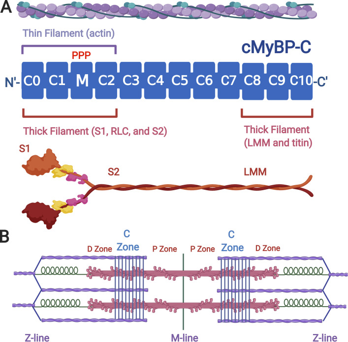
Figure 1.
cMyBP-C domain structure and binding interactions. (A) cMyBP-C (MYBPC3 gene product) is depicted as a series of blue rectangles representing either Ig- or fibronectin-like folded domains. Domains are numbered C0–C10 beginning from the N terminus to the C terminus. The position of the regulatory M-domain with three serines (PPP) that are phosphorylated by protein kinase A is shown between domains C1 and C2. Domains C0–C2 bind to thin (actin) filaments (purple) and to thick (myosin) filaments (red). Domains C8–C10 anchor MyBP-C to the thick filament by binding to the light-meromyosin (LMM) segment of myosin and to titin. (B) Cartoon of a sarcomere showing thin filaments (purple), thick filaments (red), and titin (green). MyBP-C localization is shown as a series of nine regularly spaced stripes (blue) in the C-zones of each half sarcomere. D- and P-zones of the thick filament are also indicated. Figure created with Biorender.com.

