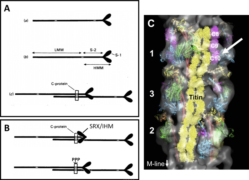
Figure 2.
Diagrams and 3-D reconstruction showing possible arrangements of binding sites for C-protein on myosin and the thick filament. (A) Diagram from Starr and Offer (1978) showing how MyBP-C binding sites could restrict myosin heads. Binding sites are indicated by the white areas in myosin tails. (A a) Separate binding sites in the light meromyosin and subfragment-2 regions. (A b) Binding site shared by the light-meromyosin (LMM) and subfragment-2 (S-2) regions. (A c) Diagram showing how in the thick filament one C-protein molecule could interact with the heavy-meromyosin (HMM) region of one myosin molecule and the LMM region of another. S-1, subfragment-1. Figure and legend reprinted from Starr and Offer (1978) with permission from Biochemical Journal. (B) Modification of myosin heads redrawn from A to represent myosin heads stabilized by MyBP-C and folded back against the thick filament in the IHM/SRX state (top) and effects of cMyBP-C phosphorylation to disrupt the IHM/SRX state (bottom). PPP, regulatory M-domain with three serines (see legend to Fig. 1). (C) 3-D reconstruction of a cardiac thick filament showing three crowns of myosin heads (1, 3, and 2), myosin S2 (red), myosin free heads (cyan), myosin blocked heads (green), titin (yellow), and MyBP-C C8-C10 domains (magenta). Possible interaction of the C10 domain of MyBP-C with a myosin free head is indicated by white arrow. Figure modified from Al-Khayat et al. (2013) with permission from Proceedings of the National Academy of Sciences of the United States of America.

