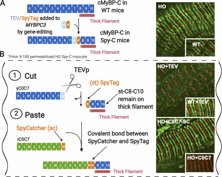
Figure 4.
Cut-and-paste approach for removal and replacement of cMyBP-C N-terminal domains in cardiomyocytes from homozygous Spy-C mice. (A) Gene-edited Spy-C mice express modified cMyBP-C with a TEVp recognition site (light blue rectangle) and a SpyTag (orange rectangle) inserted between domains C7 and C8. Inset: Immunofluorescence localization of cMyBP-C in homozygous (HO) and WT Spy-C myocytes showing expected localization (green) in each half sarcomere. Z-lines are shown stained with α-actinin (red). (B) Cut and paste of cMyBP-C in Triton X-100–permeabilized cardiomyocytes from HO Spy-C mice. (B 1) Cut: TEVp treatment releases genetically encoded (γ) C0–C7 domains, which are soluble and can be removed from sarcomeres through gentle rinsing. Inset: Immunofluorescence showing loss of cMyBP-C stripes after TEVp treatment in HO myocytes but not WT myocytes. (B 2) Paste: New recombinant rC0C7-sc domains (green; encoding any desired modification such as mutations, deletions, and fluorescent probes) are added to the bath where they become covalently attached to st-C8C10 on the thick filament via a spontaneous bond formed between SpyCatcher and SpyTag. Inset: Immunofluorescence localization showing reappearance of cMyBP-C stripes (green) after addition of rC0C7-sc in HO myocytes but not rC0C7 lacking encoded SpyCatcher in HO myocytes. Data and figure modified from Napierski et al. (2020). Figure created with BioRender.com.

