Figure 5.
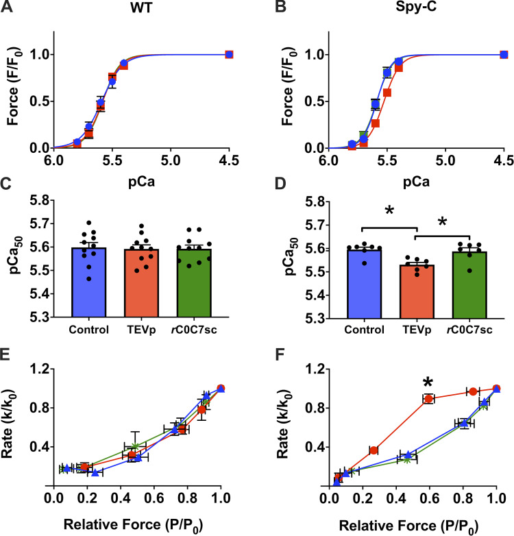
Effects of cut and paste of cMyBP-C N-terminal domains on force in skinned myocytes. (A and B) Normalized tension–pCa relationships in WT myocytes (A) or HO Spy-C myocytes (B); before TEVp treatment (control, blue), after TEVp treatment (red), and after incubation with rC0C7-sc (green). (C and D) Summary data showing average pCa50 values for each condition in WT (C) and HO Spy-C myocytes (D). Loss of C0–C7 domains after TEVp treatment caused a decrease in Ca2+ sensitivity to tension that was reversed by ligation of rC0-C7-sc in HO but not WT myocytes. (E and F) ktr data from WT (E) and HO (F) myocytes showing an increase in relative ktr at intermediate [Ca2+] after TEVp treatment that was reversed by ligation of rC0-C7-sc in HO but not WT myocytes. Data reprinted from Napierski et al. (2020).

