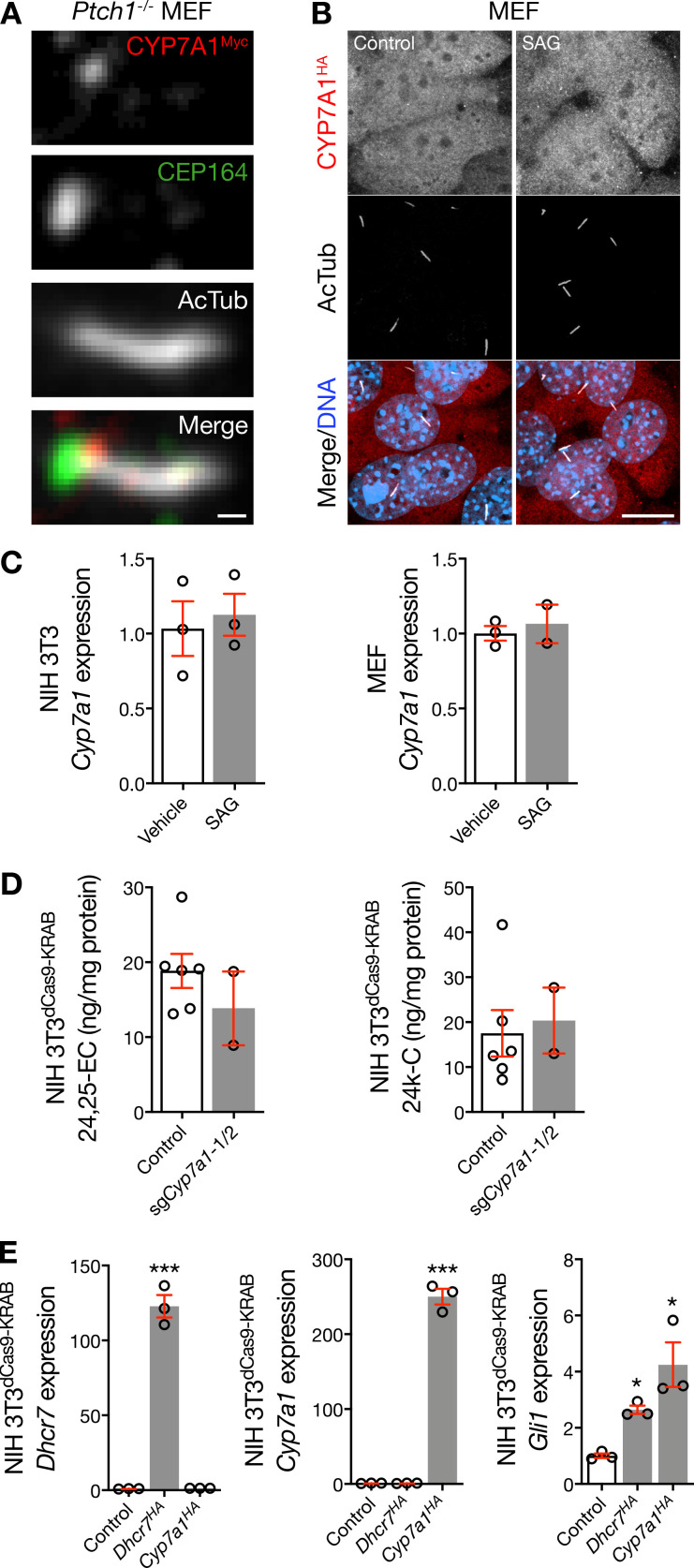
Figure S3.
CYP7A1 localizes near the ciliary base. (A) Immunofluorescence confocal microscopy of Ptch1−/− MEFs transfected with Cyp7a1Myc in pCMV6-Entry using Lipofectamine demonstrates that exogenous CYP7A1 can localize near the ciliary base, as marked by the centriole protein CEP164, after genetic Hedgehog pathway activation. Scale bar, 1 µm. (B) Immunofluorescence confocal microscopy of MEFs treated with vehicle control or SAG, and transfected with Cyp7a1HA in pCDNA3 using FuGENE, demonstrates that exogenous CYP7a1 can localize away from the ciliary microenvironment. DNA is marked with Hoechst 33342. Scale bar, 10 µm. (C) qRT-PCR assessment of NIH 3T3 cells and MEFs reveals that pharmacologic activation of the Hedgehog pathway fails to alter expression of Cyp7a1. (D) Mass spectrometry–based sterolomics of NIH 3T3dCas9-KRAB cells show that transduction of sgCyp7a1 does not alter cellular levels of Smoothened-activating oxysterols that are produced by other enzymes. (E) qRT-PCR assessment of NIH 3T3 cells demonstrates that overexpression of Dhcr7HA or Cyp7a1HA activates the Hedgehog transcriptional program (ANOVA). *, P ≤ 0.05; ***, P ≤ 0.001. Error bars represent SEM. The sample size of each experiment is represented by the number of independent data points on each graph. Each experiment is representative of at least three independent biological replicates. 24,25-EC, 24,25-epoxycholesterol; 24k-C, 24-keto-cholesterol.

