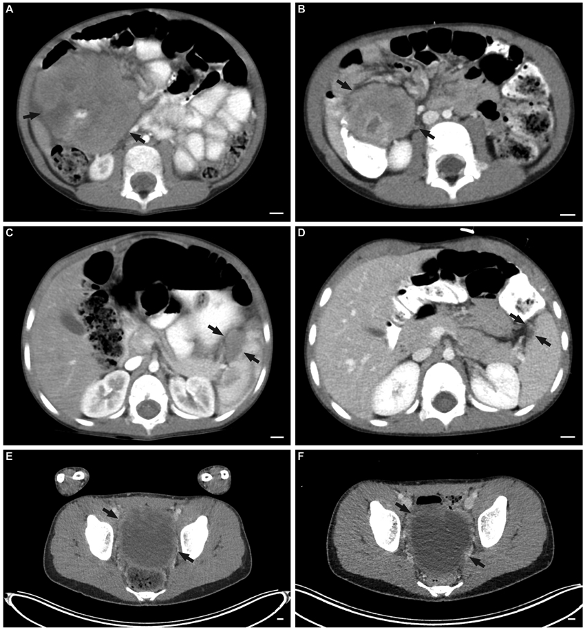
FIGURE 1.

Tumor imaging at diagnosis and after two induction cycles of VIT as per ARST08P1. Representative images of abdominal tumor (A) before and (B) after two cycles of VIT in one of the patients with partial response. Representative images of splenic lesion (C) before and (D) after two cycles of VIT in one of the patients with partial response. Representative images of a large pelvic lesion (E) before and (F) after two cycles of VIT in one of the patients with stable disease.
