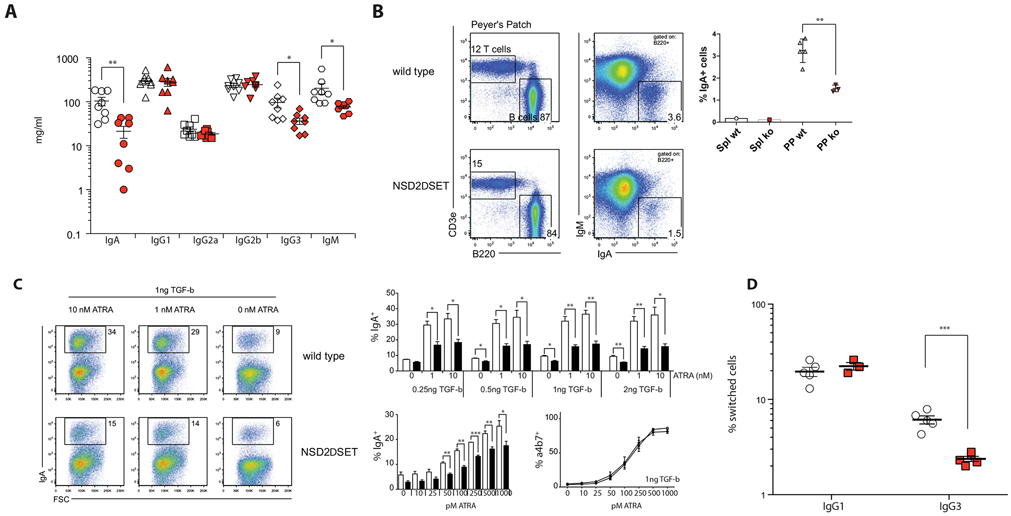
Fig. 3: NSD2 controls selective class switch recombination in B cell.

A) Concentration of serum immunoglobulins in Nsd2loxSET/loxSET (white symbols) and Mb1creNsd2loxSET/loxSET (red symbols) mice were quantified by ELISA. Each symbol represents one mouse (8 mice per genotype, 2 independent experiments). Significance was determined by unpaired t-test *: p ≤ 0.05, **: p ≤ 0.01, ***: p ≤ 0.001. B) FACS plots show relative abundance of T cells (T cells, CD3e+) and B cells (B cells, B220+); and the relative abundance of surface IgA positive (B220+IgA+) B cells in Peyer’s Patches of Nsd2loxSET/loxSET (WT) and Mb1creNsd2loxSET/loxSET (NSD2ΔSET; ko) mice. The percent of IgA+ B2 cells in spleen (Spl) and Peyer’s Patches (PP) is indicated on the right. A representative experiment out of 3 performed (3 mice each). C) NSD2ΔSET B2 cells have a defect in switching to IgA in vitro. Splenic B2 cells isolated from Nsd2loxSET/loxSET (WT) and Mb1creNsd2loxSET/loxSET (NSD2ΔSET) mice were stimulated in vitro in the presence of LPS, TGF-β and all trans-retinoic acid (ATRA). The percentage of IgA positive B cells is indicated. Representative plots of more than 3 independent experiments with 3 or more mice per group are shown. Bar diagrams indicate the percent of IgA positive B2 cells after 3 days in various culture conditions. Control Nsd2loxSET/loxSET cells (white bars) and cells from Mb1creNsd2loxSET/loxSET (black bars) mice (top) are shown. NSD2ΔSET B2 cells have a defect in switching to IgA, but not in the induction of the integrin α4β7 in response to ATRA. The percentage of IgA positive B cells is response to LPS and a stable amount of TGF-β (1ng) with increasing amounts of ATRA (0-1000pM) in Nsd2loxSET/loxSET (white bars) and Mb1creNsd2loxSET/loxSET (black bars) B2 cells is indicated (bottom left). The percentage of integrin α4β7-positive B cells is response to LPS and a stable amount of TGF-β (1ng) with increasing amounts of ATRA (0-1000pM) in Nsd2loxSET/loxSET (open symbols) and Mb1creNsd2loxSET/loxSET (closed symbols) B2 cells is indicated (bottom right). D) NSD2ΔSET B2 cells have a defect in switching to IgG3, but not IgG1 in vitro. B2 cells isolated from Nsd2loxSET/loxSET (open symbols) and Mb1creNsd2loxSET/loxSET (red symbols) mice were stimulated in vitro in the presence of LPS or LPS+IL4. The percentages of switched B cells after 3 days in vitro culture are indicated. Each symbol represents one mouse (3-5 mice per genotype, 2 independent experiments). Significance was determined by unpaired t-test *: p ≤ 0.05, **: p ≤ 0.01, ***: p ≤ 0.001.
