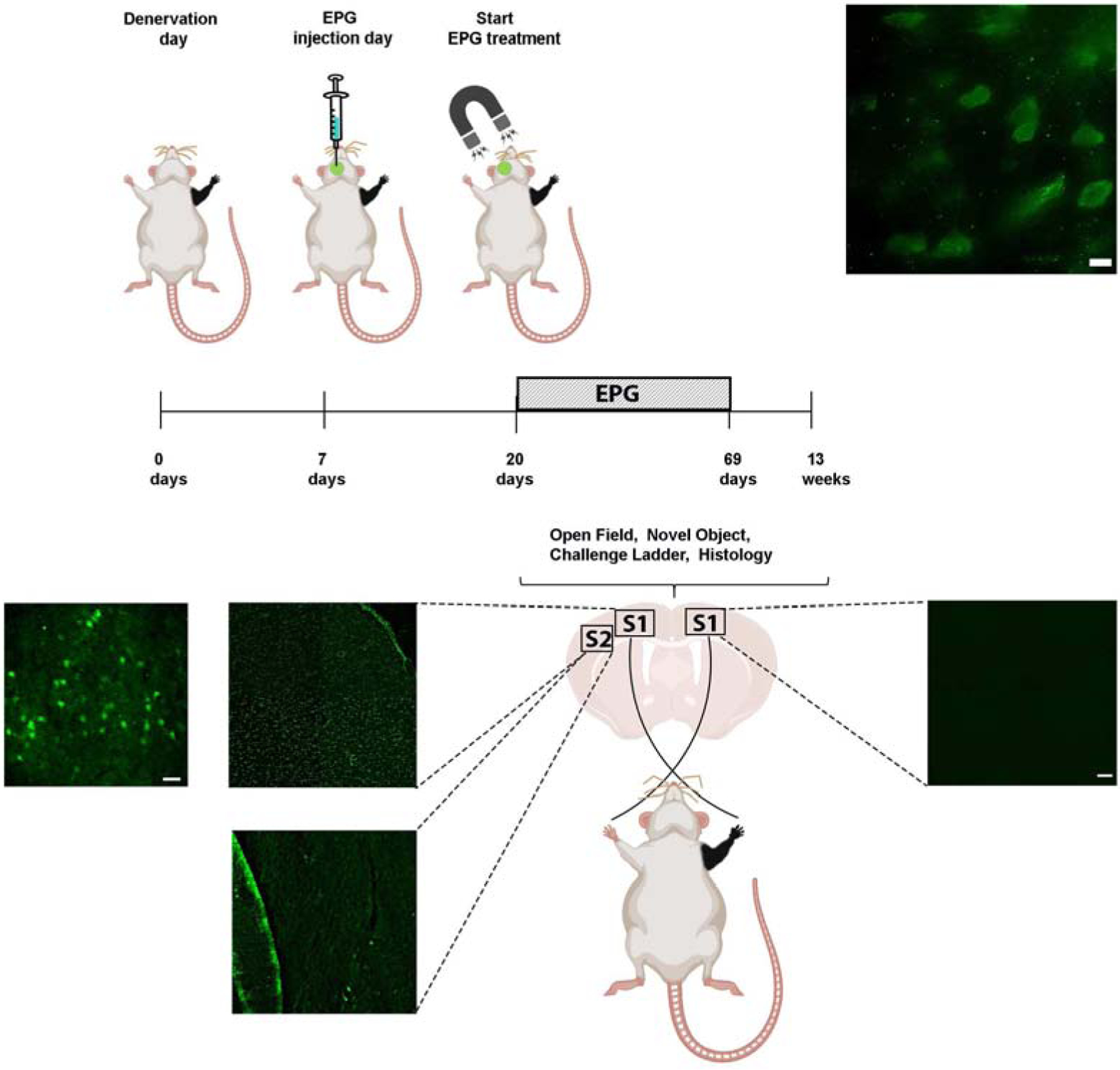
Fig. 5.

Diagram demonstrating the experimental design of neuromodulation via EPG. Virus encoding to the EPG was stereotaxicly injected into the left S1, contralateral to the denervated forepaw (Den-EPG). Denervated control rats were injected with a virus encoding for a fluorescence protein (Den-Control). An electromagnet was placed over the left S1 starting three weeks following stereotaxic injection. The electromagnet delivered magnetic field stimulation for 16 minutes once a day, for 30 days. Immunostaining images in the primary somatosensory cortex showing EPG expression in fixed brain sections using anti-FLAG antibody in left S1, and right, non-injected S1. On the left S1, EPG can be detected with high-magnification of 100× (upper panel, scale bar= 10 μm), 40× (Scale bar=20 μm) and 4× magnification (Scale bar = 50 μm). No EPG was detected in secondary somatosensory cortex (S2), and in the right, non-injected S1. 4× magnification (Scale bar = 50 μm). No EPG was detected in secondary somatosensory cortex (S2), and in the right, non-injected S1.
