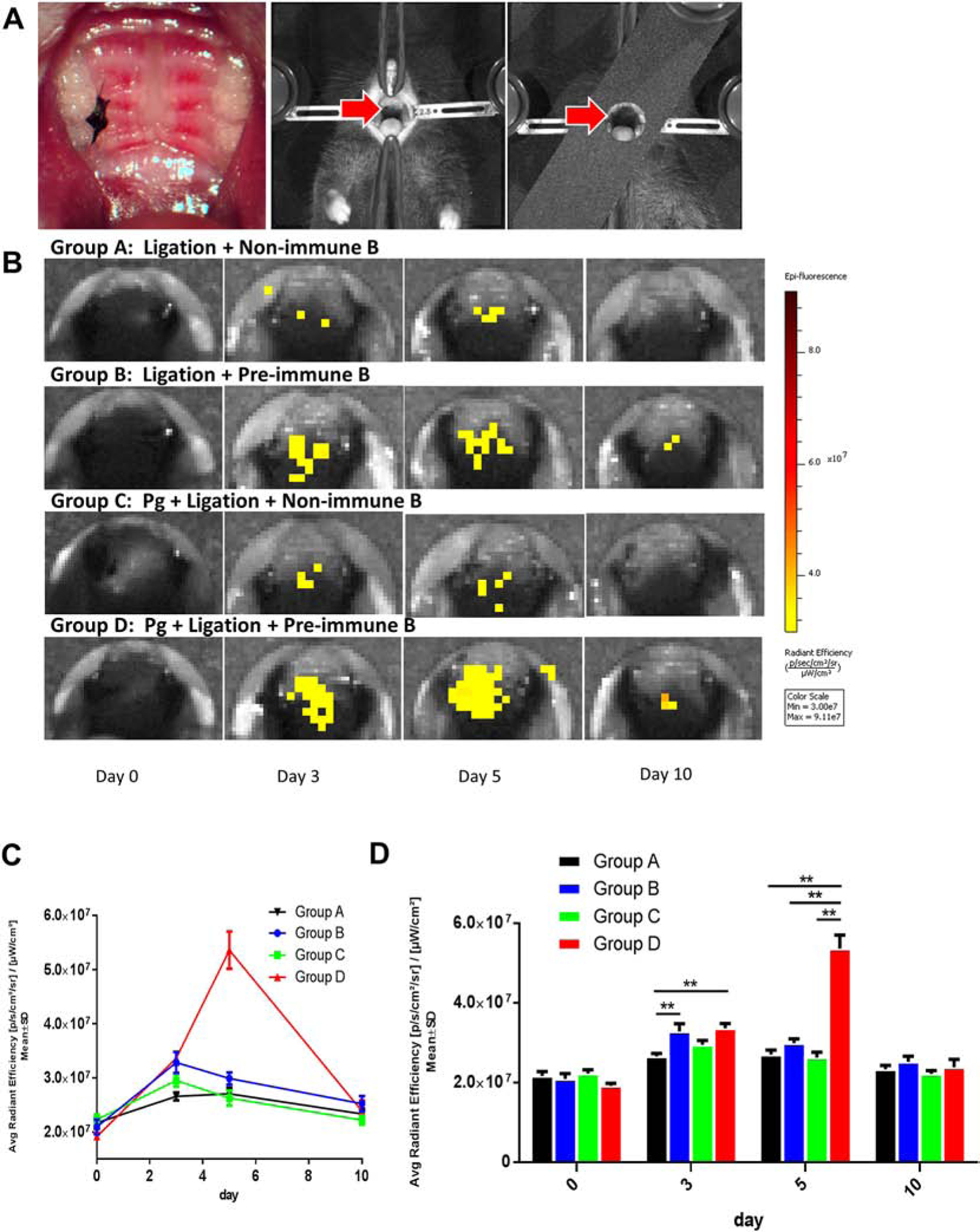
Figure 3. In-vivo imaging revealed antigen-directed gingival B10 infiltration in experimental periodontitis.

A. Silk ligature was placed around the right second maxillary molar and mice were positioned in acquisition platform in IVIS Spectrum in vivo imaging system. B. In vivo fluorescence showed that the antigen-specific B cells could be recruited more efficiently on day 3 in group B and D, comparing with that in group A and C, which were transferred with non-specific B cells. C. The level of fluorescence intensity showed a significant peak on day 5 in group D, comparing with the other 3 groups. D. On day 3, the fluorescence of group D was significantly higher than that of group A and B. While on day 5, the fluorescence of group D was significantly higher than those of the other 3 groups. (Mean ± SEM, n=5, *p < 0.05, **p < 0.01)
