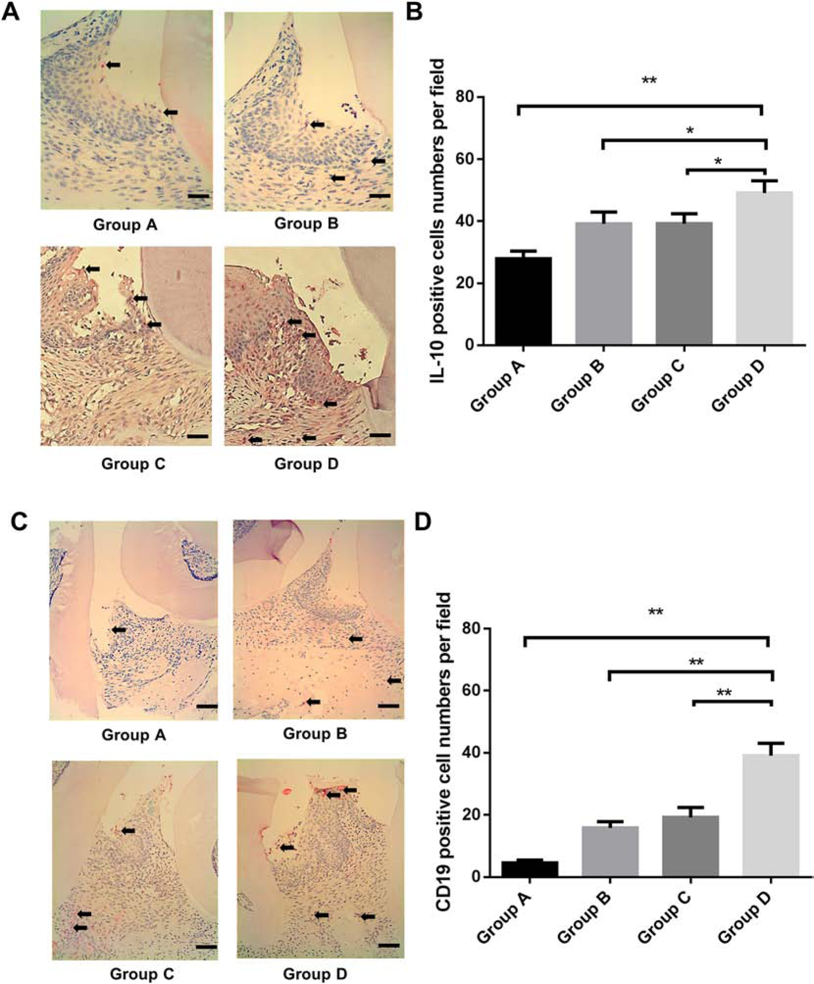
Figure 4. Gingival IL-10 expression levels in all experimental periodontitis groups.

A. Immunohistochemistry staining of IL-10 in the gingival tissue around ligation site were performed in Group A (ligation + non-immunized B cells), Group B (ligation + pre-immunized B cells), Group C (P. gingivalis ligation + non-immunized B cells) and Group D (P. gingivalis ligation + pre-immunized B cells) (Scale bar, 100 μm). B, the IL-10 positive cell numbers per field was analyzed and compared between groups. (Mean ± SEM, n=5, *p < 0.05, **p < 0.01). C. Immunohistochemistry staining of CD19 positive cells in the gingival tissue around ligation site were performed in Group A, B, C, and D. (Scale bar, 200 μm). D, The CD19 positive cell numbers per field was analyzed and compared between groups. (Mean ± SEM, n=5, **p < 0.01).
