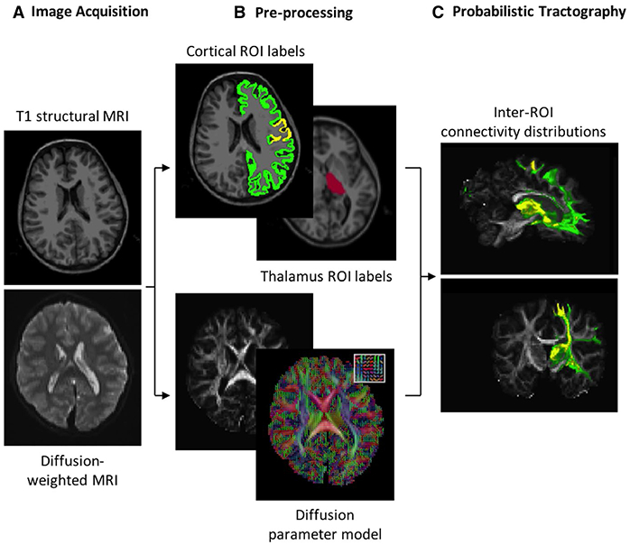
FIGURE 1.

Thalamocortical tractography processing pipeline. A, High-resolution structural and diffusion magnetic resonance images (MRIs) are acquired. B, The Desikan-Killiany atlas is used to label Rolandic (yellow) and non-Rolandic (green) cortex and the thalamus (red) in structural MRIs (top). Diffusion MRIs are used to extract diffusion parameters per voxel from 64 gradient directions (example principal directions of diffusion for 25 voxels shown in inset). C, Distribution of diffusion parameters is repeatedly sampled to infer the probability of white matter tracts between regions of interest (ROIs)
