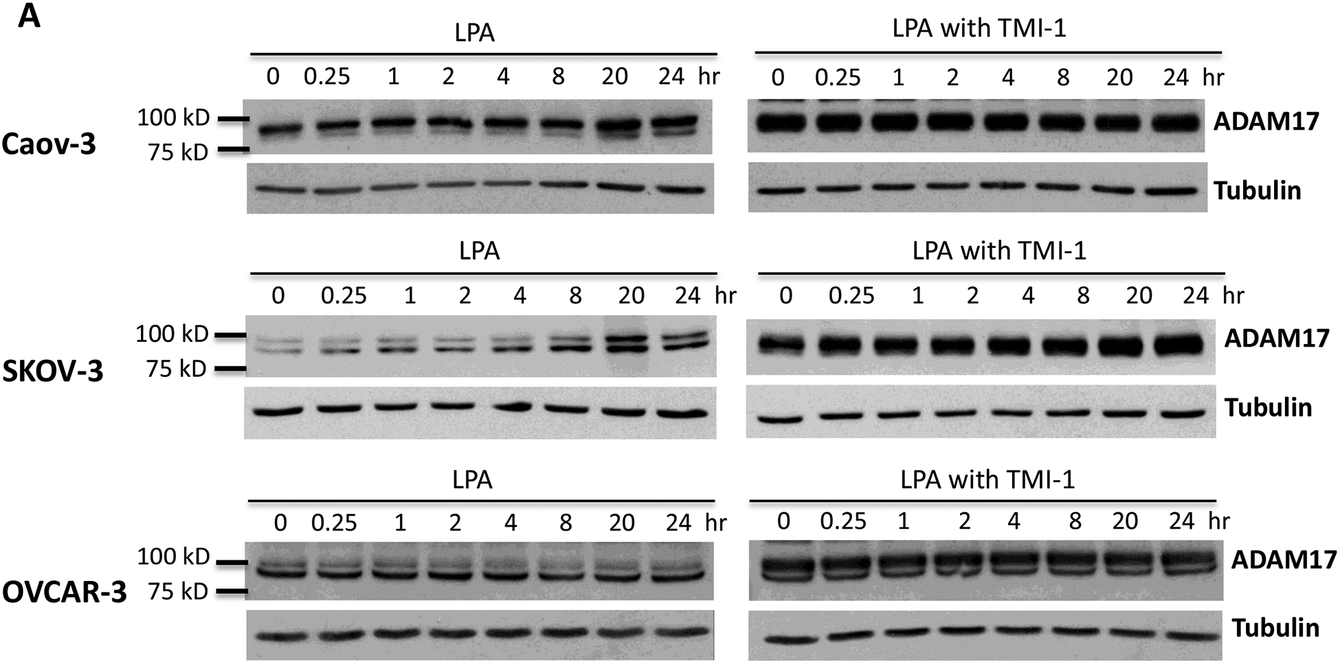
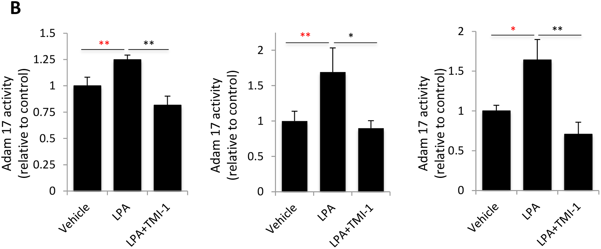
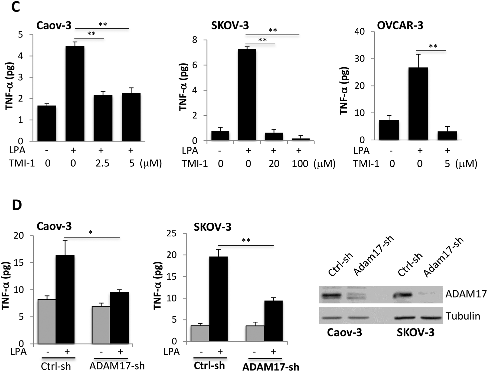
Figure 3. LPA-driven TNF-α release relies on the activity of ADAM17.


A. Caov-3, SKOV-3 and OVCAR-3 cells were treated with 10 μM LPA for the indicated periods of time with or without TMI-1. TMI-1 was added at 2.5, 20 and 5 μM to Caov-3, SKOV-3 and OVCAR-3, respectively, 1 hour before LPA treatment. Expression of ADAM17 was assessed by immunoblotting analysis. B. Ovarian cancer cell lines in 24-well plated were serum-starved and treated with LPA (10 μM) with or without MTI-1 (5 μM for Caov-3 or OVCAR-3 and 20 μM for SKOV-3) for 30 minutes before addition of the quenched fluorogenic substrate for measurement at 37 °C for 1 hour as detailed in Materials and Methods. The results were presented as fold changes in activity relative to the un-stimulated control cells (defined as 1 fold). C. The ovarian cancer cell lines in 12-well plates were stimulated with LPA in the presence of indicated concentrations of TMI-1. TNF-α levels in culture supernatants were quantified with ELISA and presented as in Fig. 1A. D. ADAM17 was silenced in Caov-3 and SKOV-3 cells by lentivirally-mediated shRNA to determine the effect of ADAM17 deficiency on LPA-induced TNF-α release. ADAM17 knockdown efficiency was confirmed by immunoblotting analysis.
