
Figure 6. LPA-induced TNF-α is functionally sufficient to reinforce IL-8, IL-6 and CXCL1 production.
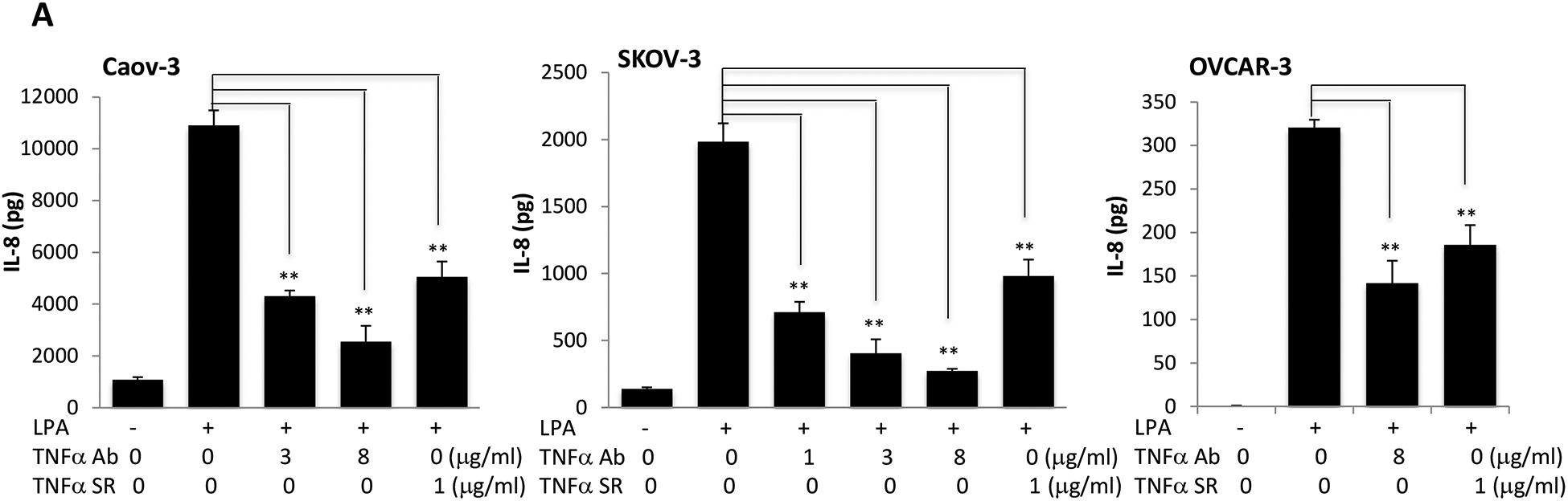
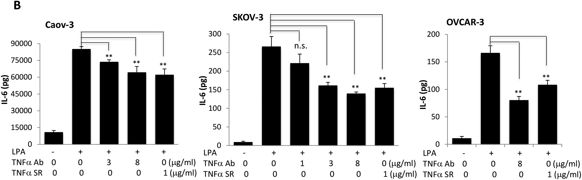
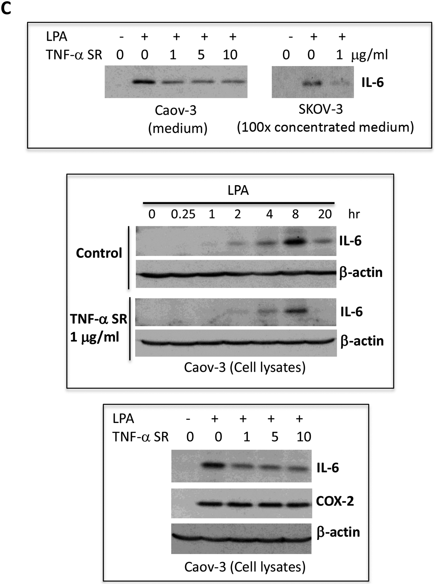
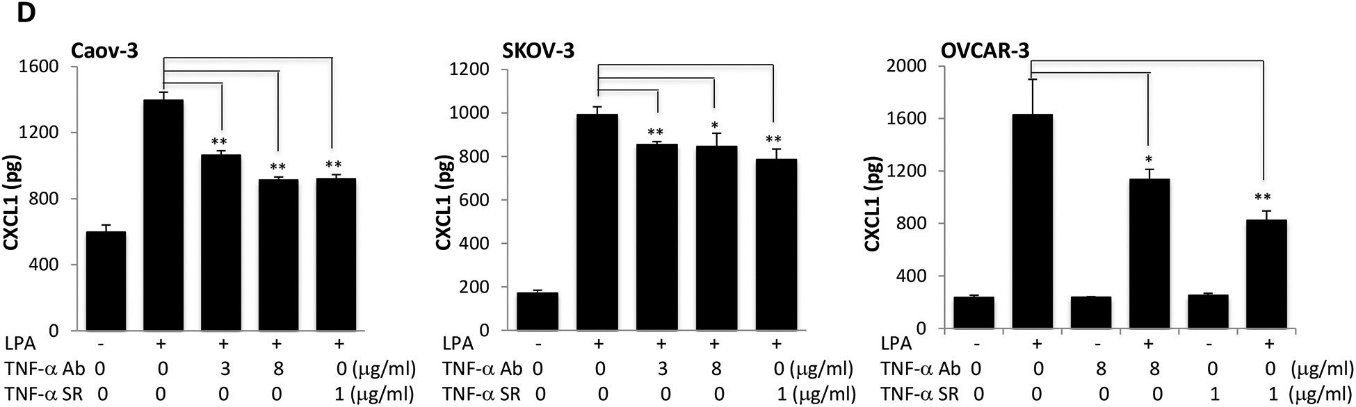
Caov-3, SKOV-3 and OVCAR-3 cells were treated for 24 hours with LPA as in Fig. 1 in the presence of vehicle, TNF-α Ab or TNF-α SR at the indicated concentrations. These TNF-α blocking agents were added simultaneously with LPA. Concentrations of IL-8 (A), IL-6 (B), CXCL1 (D) in culture supernatants were measured with ELISA and the results presented in the same way as TNF-α described in Fig. 1A. C. IL-6 protein in culture supernatants of Caov-3 cells or in 100X concentrated supernatants of SKOV-3 cells were assessed by immunoblotting analysis (upper). The time-dependent induction of intracellular IL-6 in the presence or absence of TNF-α SR was examined by immunoblotting analysis of cellular lysates (middle). The specificity of TNF-α SR inhibition of IL-6 was verified by lack of an inhibitory effect on cellular COX-2, another LPA-induced gene (lower).