
Fig. 3. Knockdown of Inner-Ring NUPs Disrupts Cilia.
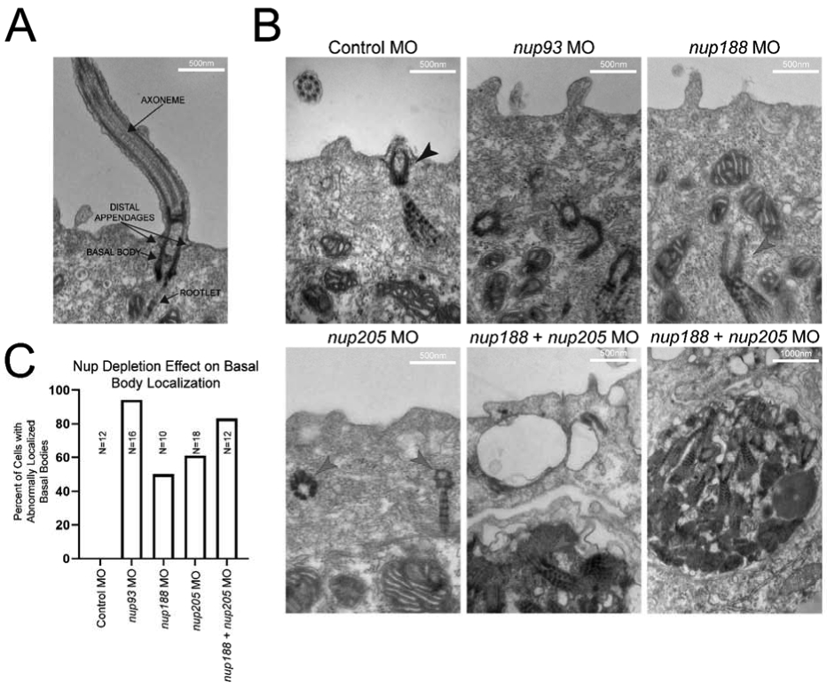
(A) Examples of cilia components. (B) Representative images of cilia within MCCs of control and inner-ring NUP knockdown morphant embryo epidermis. Black arrowheads indicate properly positioned ciliary components, red arrowheads indicate mispositioned ciliary components beneath the cell surface. Note the apparent inclusion body with multiple ciliary components in the cases of nup188 + nup205 knockdown. (C) Percentages of visualized cells with abnormally positioned basal bodies at cell surface for each condition (basal bodies within 100nm of surface were considered to be normally localized). Ns indicate number of cells assessed from a single embryo per condition.