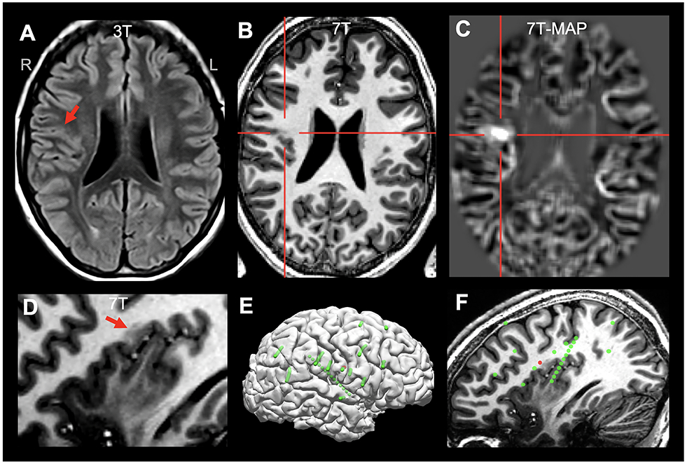
Figure 2.

Illustration of concordant ICEEG onset and 7T finding (detected by MAP-guided review) in a patient with a subtle FCD in the right parietal operculum (highlighted by red arrows or crosshairs), characterized by gray-white blurring and T1-weighted signal abnormality. A: 3T FLAIR (axial) on which the lesion was difficult to appreciate; B: 7T T1-weighted MP2RAGE (axial); C: 7T MAP junction file (axial); D: 7T MP2RAGE (sagittal, zoomed in); E: all implanted SEEG electrodes (green spheres: all implanted electrodes; red spheres: ictal onset); F: sagittal view with 7T finding and SEEG electrode location; the 7T-MAP-detected FCD lesion was concordant with ictal onset shown by the SEEG. The patient was seizure-free at 1 year follow up, following complete resection of the lesion. Histopathology was not available due to fragmented tissue.
