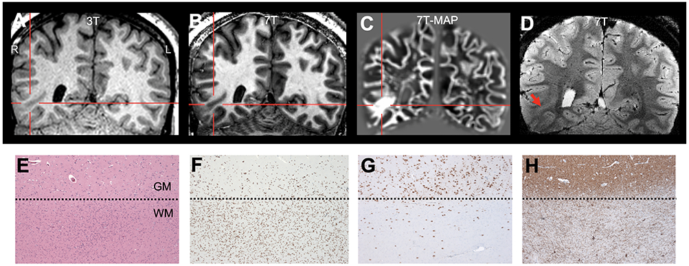
Figure 5.

Illustration of 7T findings in a patient with mild malformation of cortical development with oligodendroglial hyperplasia (MOGHE) in the right basal tempo-occipital region. A: 3T T1-weighted MPRAGE (coronal); B: 7T T1-weighted MP2RAGE (coronal); C: 7T MAP junction file (coronal); D: 7T T2*-weighted GRE (coronal). In this patient, the lesion was detected by MAP-guided 7T review. The patient had seizure recurrence (although at a reduced seizure frequency) at 1 year following partial resection of the abnormality. Pathology panel E: increased cell densities at the gray-white-matter junction, with patchy increased cellularity (H&E staining). F: Olig2 immunohistochemistry of the same area as A proving the oligodendroglial origin of cells. In addition to the oligodendroglial hyperplasia, NeuN (G) and Map2 (H) immunohistochemical stainings demonstrated blurred gray-white-matter boundaries with increased density of heterotopic neurons in the subjacent white matter. Dotted lines in E-H: gray-white-matter junction. Scale bar in E = 200μm, which also applies to F-H.
