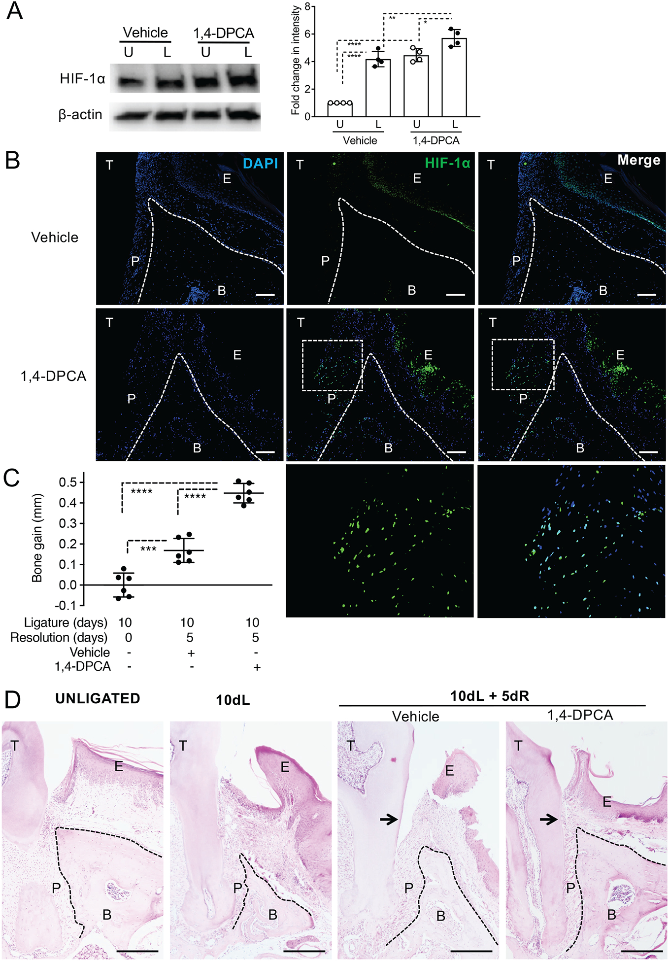
Figure 1: 1,4-DPCA/hydrogel promotes HIF-1α protein levels in the periodontium and alveolar bone regeneration.

Mice were subjected to LIP for 10 days followed by 5 days without ligatures to enable resolution. At the onset of resolution (day 10), the mice were subcutaneously injected with 1,4-DPCA/hydrogel, or vehicle alone, and were euthanized at day 15 for analysis. (A) The expression of HIF-1α in gingival tissue lysates was analyzed by immunoblotting (left panel). The relative intensity of immunoblotting signals of HIF-1α was quantified by densitometry (right panel). U, unligated; L, ligated. (B) Periodontal tissue sections were stained for HIF-1α protein (green) and DAPI (blue). Scale bars, 100 μm. Bottom row images are magnifications of the rectangle-demarcated areas of the images above. (C) Bone heights (CEJ-ABC distance) were measured and CEJ-ABC data were transformed to indicate bone gain, relative to the bone heights at day 10 (baseline). (D) Periodontal tissue sections were stained with hematoxylin and eosin. Scale bars, 500 μm. Black arrows indicate increased attachment gain in 1,4-DPCA/hydrogel group as compared to vehicle control. NL, not ligated; 10dL, 10 days ligated; 10dL + 5dR, 10 days ligated and 5 days with ligatures removed (resolution). B, Bone; E, Epithelium; P, Periodontal ligament; T, Tooth. Data are shown as means ± SD (A, n=4 mice/per group; B, n=6 mice/per group from two independent experiments, each performed in triplicates). *P < 0.05, **P < 0.01, ***P < 0.001, ****P < 0.0001 (ANOVA with Turkey’s multiple-comparisons test).
