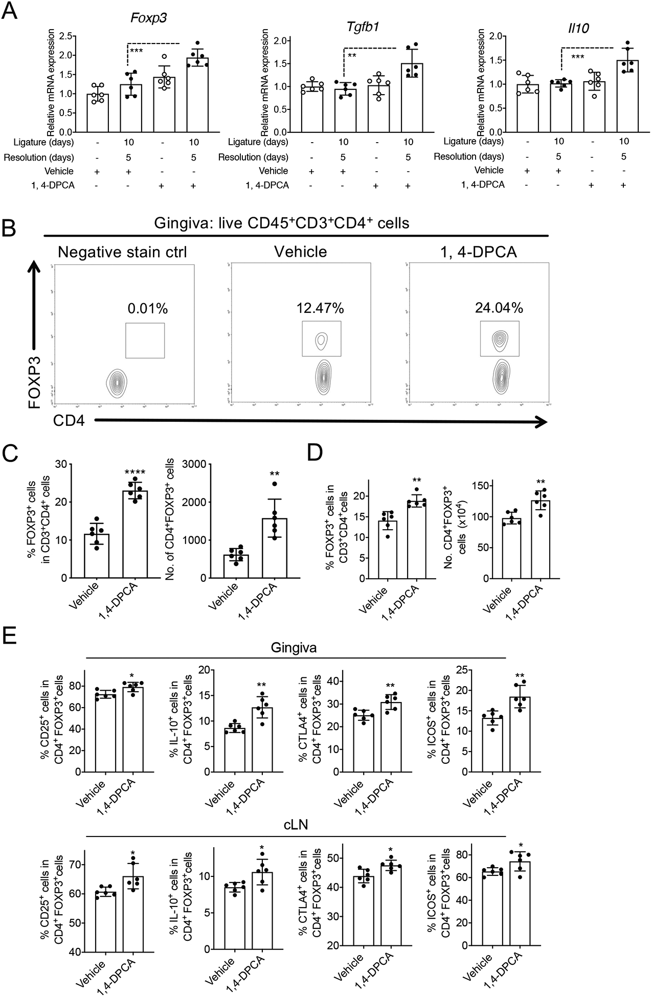
Figure 3: Treatment with 1,4-DPCA/hydrogel leads to increased number of Tregs during the resolution of periodontitis.

Mice were subjected to LIP for 10 days followed by 5 days without ligatures to enable resolution. At the onset of resolution (day 10), the mice were subcutaneously injected once with 1,4-DPCA/hydrogel, or vehicle alone, and were euthanized on day 15 for analysis. (A) Gene expression in the gingival tissue was analyzed by quantitative real-time PCR. The mRNA expression of the indicated molecules was normalized to that of Hprt; results are presented as fold change relative to the mRNA levels of the unligated contralateral sites of the vehicle control-treated mice, assigned an average value of 1. (B) Representative FACS plots of Treg cells and (C) bar graphs showing percentage of Treg cells in CD4+ T cells (left) and their absolute numbers (right), in gingival tissues of vehicle control-treated and 1,4-DPCA/hydrogel-treated mice on day 15. (D) Bar graphs showing percentage of Treg cells in CD4+ T cells (left) and their absolute numbers (right), in cervical lymph nodes (cLNs) of vehicle control-treated and 1,4-DPCA/hydrogel-treated mice on day 15. (E) Frequency of CD25+, IL-10+, CTLA-4+ and ICOS+ in CD4+FOXP3+Treg cells in gingiva (top) and cLNs (bottom) of vehicle control-treated and 1,4-DPCA/hydrogel-treated mice on day 15. Data are shown as means ± SD (n=6 mice/per group from two independent experiments, each performed in triplicates). *P < 0.05, **P < 0.01, ***P < 0.001, ****P < 0.0001. (Student’s t-test).
