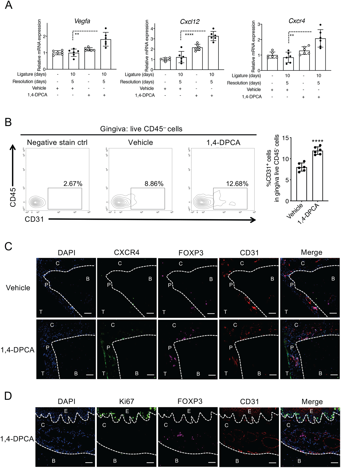
Figure 4: 1,4-DPCA/hydrogel promotes angiogenesis and the emergence of Treg cells in and around vessels of the periodontium during the resolution of periodontitis.

Mice were subjected to LIP for 10 days followed by 5 days without ligatures to enable resolution. At the onset of resolution (day 10), the mice were subcutaneously injected once with 1,4-DPCA/hydrogel or vehicle alone and were euthanized on day 15. (A) Gingival tissues were analyzed for gene expression by quantitative real-time PCR. The mRNA expression of the indicated molecules was normalized to that of Hprt and presented as fold change relative to the mRNA levels of the unligated contralateral sites of the vehicle control-treated mice, assigned an average value of 1. (B) Gingival tissues were harvested on day 15 and processed for flow cytometric analysis. The cells obtained were stained and gated for live CD45−CD31+ cells; shown are the percentage of CD31+ cells in CD45− cells. (C) Periodontal tissue sections were stained for CXCR4 (green), FOXP3 (purple), CD31 (red), and DAPI (blue). Scale bars, 100 μm. (D) Periodontal tissue sections were stained for Ki67 (green), FOXP3 (purple), CD31 (red), and DAPI (blue). Scale bars, 20 μm. B, Bone; C, Connective tissue; E, Epithelium; P, Periodontal ligament; T, Tooth. Data are shown as means ± SD (n=6 mice/per group from two independent experiments, each performed in triplicates). **P < 0.01, ****P < 0.0001 (Student’s t-test).
