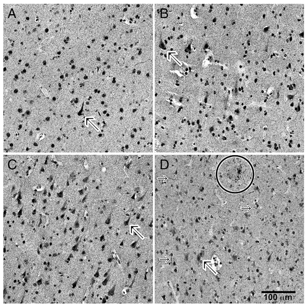
FIGURE 1:

Immunohistochemical labeling of sections for pS616IRS1. Photomicrographs of layer III of middle frontal gyrus cortex immunohistochemically labeled for pS616IRS1 are shown. (A) A 90-year-old woman without cognitive impairment or diabetes. (B) A 84-year-old man without cognitive impairment but with diabetes. (C) A 86-year-old woman with cognitive impairment and without diabetes. (D) A 90-year-old woman with cognitive impairment and diabetes. In all photomicrographs, note frequency of nonnuclear cytoplasmic immunolabeling (diagonal arrows in each of A–D, and especially in D). Also note immunolabeled neuritic threads (horizontal arrows) and neuritic plaque (circle).
