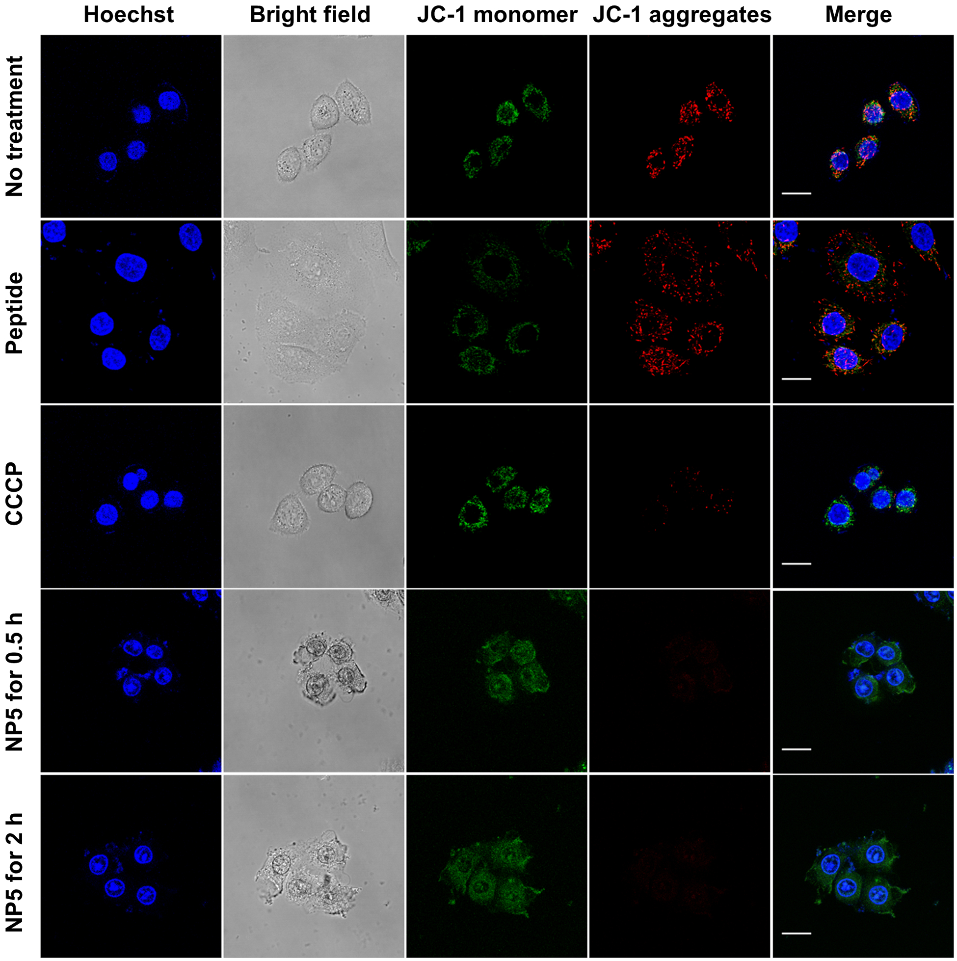
Figure 5.

Assessment of mitochondrial dysfunction induced by the peptide-containing materials using JC-1 probe. Live-cell confocal microscopy images of HeLa cells incubated with KLA peptide, CCCP, NP5 for desired periods of time. Prior to imaging, cells were stained with 2 μM of JC-probe (green, monomer, λex/em = 488 nm/510–550 nm; red, J-aggregates, λex/em = 488 nm/585–649 nm) and then Hoechst 33342 (blue, λex/em = 405 nm/ 420–480 nm). Scale bars, 20 μm.
