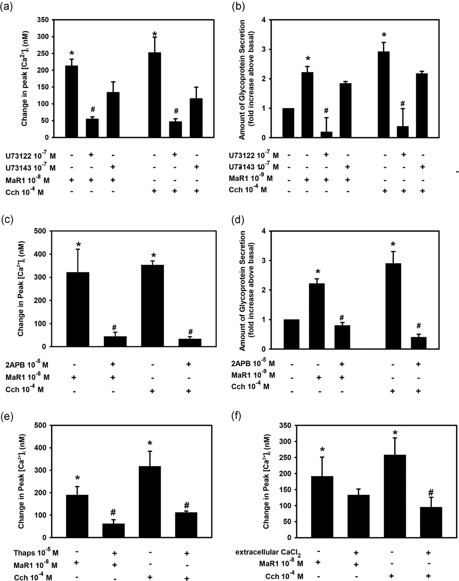
FIGURE 4.

Inhibition of phospholipase C (PLC) blocks maresin 1 (MaR1)-stimulated increase in [Ca2+]i. Goblet cells were incubated with the PLC inhibitor U73122 (10−7 M), its negative control U73143 (10−7 M, a and b) or (c and d) IP3 receptor inhibitor 2-APB (10−5 M) for 30 min and stimulated with MaR1 at 10−8 M for [Ca2+]i or 10−9 M for secretion or carbachol (Cch, 10−4 M). (a) and (c) show the change in peak [Ca2+]i while (b) and (d) show high molecular weight glycoprotein secretion. (e) Rat conjunctival goblet cells were preincubated with the SERCA-inhibitor thapsigargin (10−5 M) for 15 min and stimulated with either MaR1 (10−8 M) or carbachol (Cch, 10−4 M). (f) Goblet cells were incubated with KRB with or without CaCl2 and stimulated with MaR1 (10−8 M) or Cch (10−4 M). (e and f) The change in peak [Ca2+]i. Data are mean ± SEM of three (a–e) or five (f) experiments. *Significance above basal. #Significant difference from MaR1 or Cch alone. 2-APB, 2-aminoethoxydiphenyl borate; [Ca2+]i, intracellular Ca2+; KRB, Krebs–Ringer bicarbonate buffer; SEM, standard error of the mean; SERCA, sarco/endoplasmic reticulum
