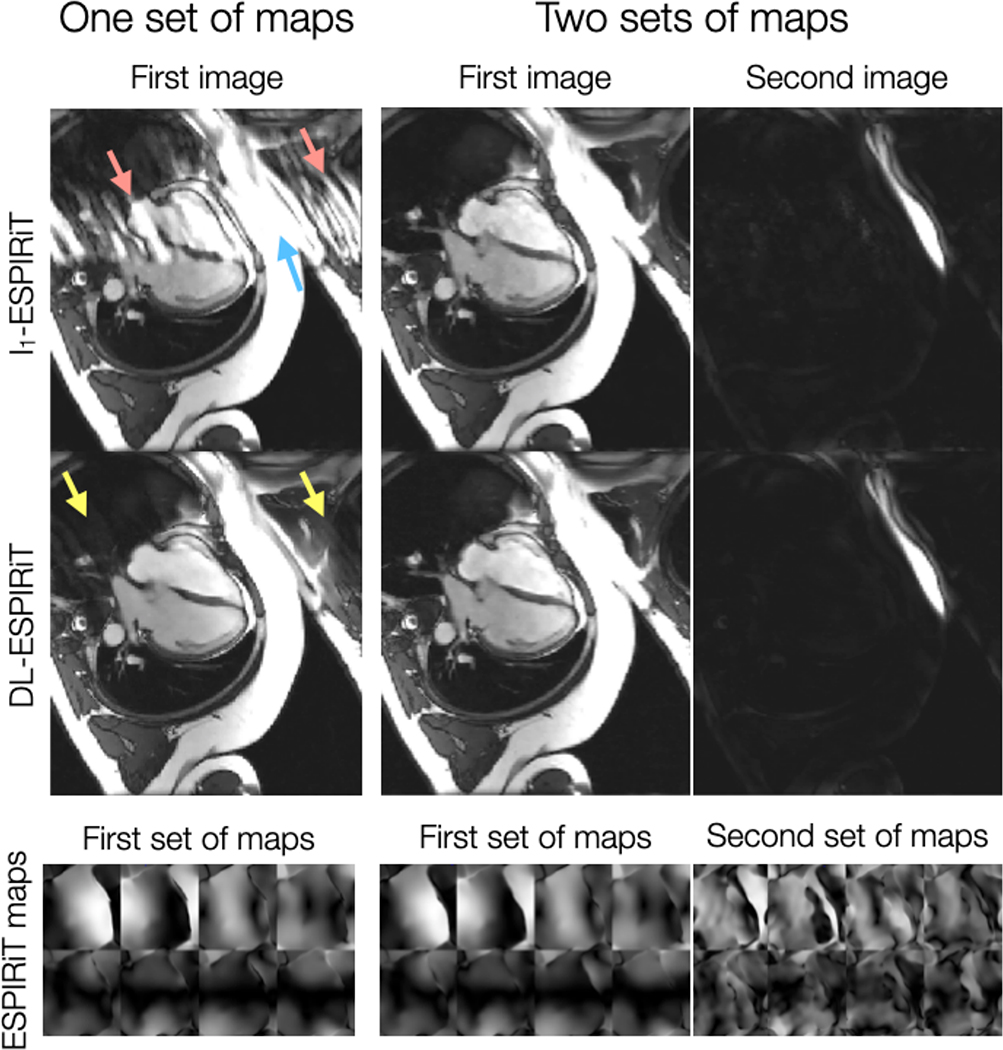
Figure 3.
A fully-sampled 2D cardiac cine acquisition in the standard 4-chamber view is retrospectively undersampled to simulate 10X acceleration, and then reconstructed using l1-ESPIRiT and (2+1)D DL-ESPIRiT with one and two sets of ESPIRiT maps. Anatomy overlap is present along the chest wall (blue arrow), which causes significant ghosting along the phase encoding direction of the single-set l1-ESPIRiT reconstruction (red arrows). Some ghosts are present in the single-set (2+1)D DL-ESPIRiT reconsruction (yellow arrows), but they are largely reduced compared to the single-set l1-ESPIRiT reconstruction. Both double-set reconstructions are able to capture overlapping anatomies, separate them into two complex-valued channels (one for each set of maps), and completely eliminate ghosting artifacts. A corresponding video of these images is shown in Supporting Information Video S1.

