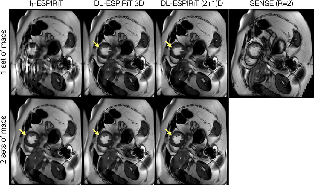
Figure 4.
With IRB approval, a prospectively undersampled (R=12) dataset is acquired from a pediatric patient within a single breath-hold on a 1.5T scanner. The images shown are reconstructed using l1-ESPIRiT, 3D DL-ESPIRiT, and (2+1)D DL-ESPIRiT algorithms with one and two sets of ESPIRiT maps. For reference, a standard cardiac cine image acquired with 2-fold uniform undersampling (R=2) and reconstructed using SENSE is shown in the top right. Subtle motion artifacts arising from respiratory motion are shown in this image because the patient had difficulty holding their breath. Anatomy overlap is present along the chest wall, which causes severe aliasing on top of the heart as shown in the l1-ESPIRiT reconstruction with one set of sensitivity maps. This artifact manifests as blurring in DL-ESPIRiT reconstructions with one set of ESPIRiT maps, and is most evident along left ventricle papillary muscles (yellow arrow). Both l1-ESPIRiT and DL-ESPIRiT reconstructions resolve these artifacts when two sets of ESPIRiT maps are used. Corresponding videos for each reconstruction are shown in Supporting Information Video S3.

