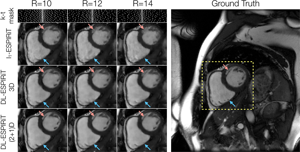
Figure 6.
A fully-sampled dataset acquired on a 3.0T scanner is retrospectively undersampled by factors of 10, 12, and 14 using variable density masks. As expected, reconstructed mid-systolic frames become progressively blurrier as the acceleration rate is increased. This is particularly evident in smaller structures such as left ventricular trabeculations (red arrow). However, the (2+1)DM2 DL-ESPIRiT network retains sharpness of this structure as the acceleration rate is increased. However, none of the reconstruction methods were able to recover tiny papillary muscle structures inside of the right ventricle for this range of acceleration rates (blue arrows). Corresponding videos of reconstructions at each acceleration rate shown here are shown in Supporting Information Video S5.

