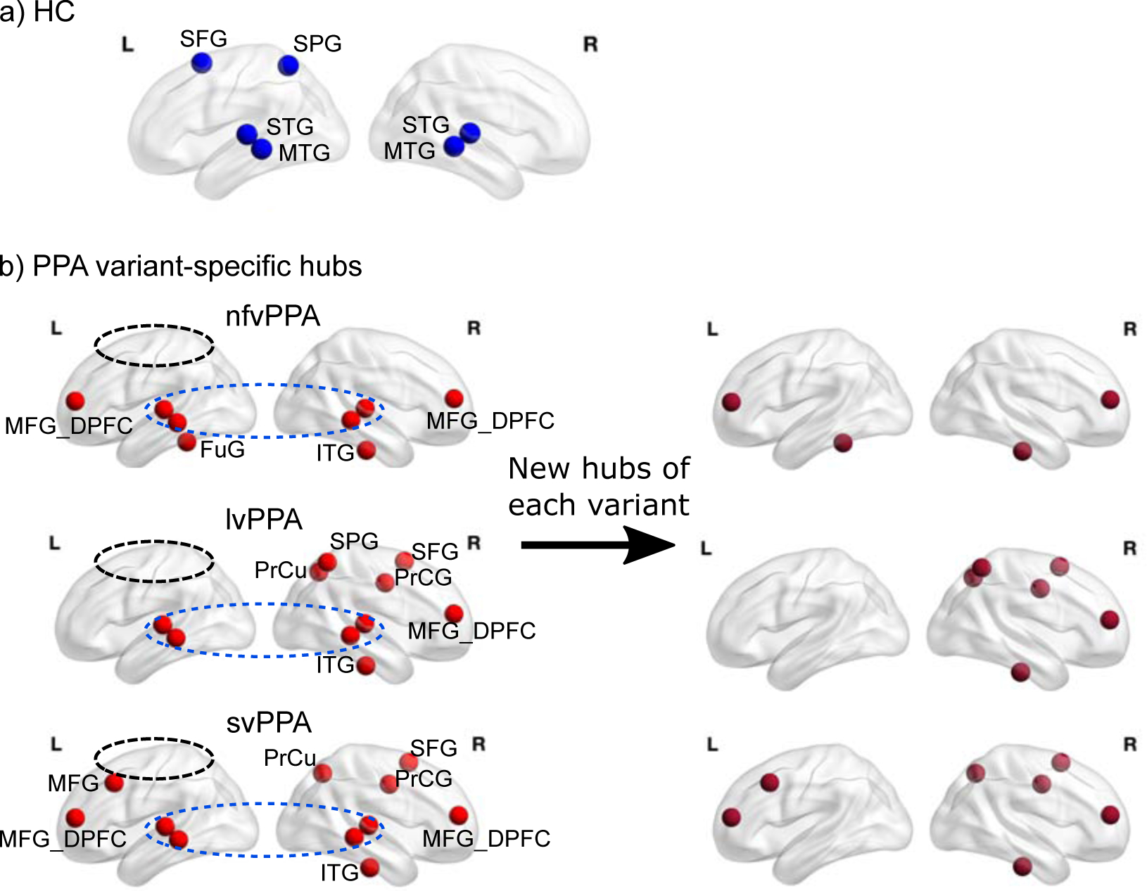
Figure 3. Connector hub distributions.

a) Connector hubs identified in the healthy control (HC) group which correspond to ROIs with the highest participation coefficient (PC) scores across the whole brain (>1SD above the mean). b) Connector hubs identified for each variant group, top to bottom: non-fluent (nfvPPA), logopenic (lvPPA), semantic (svPPA). Compared to the HC, all three variants “lost” the left SFG and the left SPG (circled) and the bilateral S/MTG are retained (circled). On the right panel the new hubs (present in PPA but not in the HC) for each variant are shown. SFG: superior frontal gyrus, SPG: superior parietal gyrus, S/MTG: superior/middle and temporal gyri, MFG: middle frontal gyrus, MFG_DPFC: middle frontal dorsal prefrontal cortex. ITG: inferior temporal gyrus, FuG: fusiform gyrus, PrCG: precentral gyrus, PrCu: precuneus.
