
Figure 3.
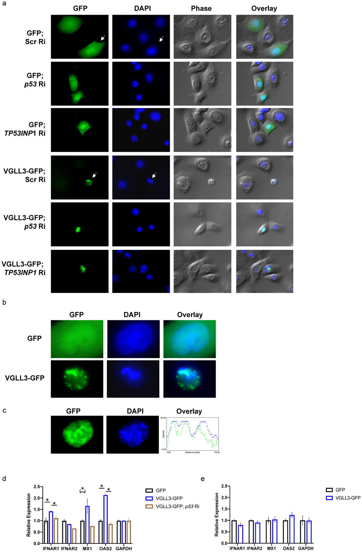
VGLL3 excess causes regulated, inflammatory cell death. a, keratinocyte cell morphology (phase) and DNA status (DAPI) upon transfection with indicated constructs in female keratinocytes. Ri, RNAi. Scr, Scrambled. Zoom-in pictures of arrow-pointed cells are shown in b to demonstrate chromatin condensation. c, overlap between GFP and DAPI signals in female keratinocyte expressing VGLL3-GFP. d, qRT-PCR of indicated genes in THP-1 cells migrated to conditioned media from female keratinocytes overexpressing GFP, VGLL3-GFP, and VGLL3-GFP with siRNA against p53. n=3. e, qRT-PCR of indicated genes in female keratinocytes overexpressing GFP and VGLL3-GFP. n=3. Mean ±s.e.m, * P < 0 .05, (*) P < 0.1, Student’s t-test.