
Figure 2. Confirmation that Noise Exposure Causing Hearing Loss Unbalances the Cochlear Proteome.
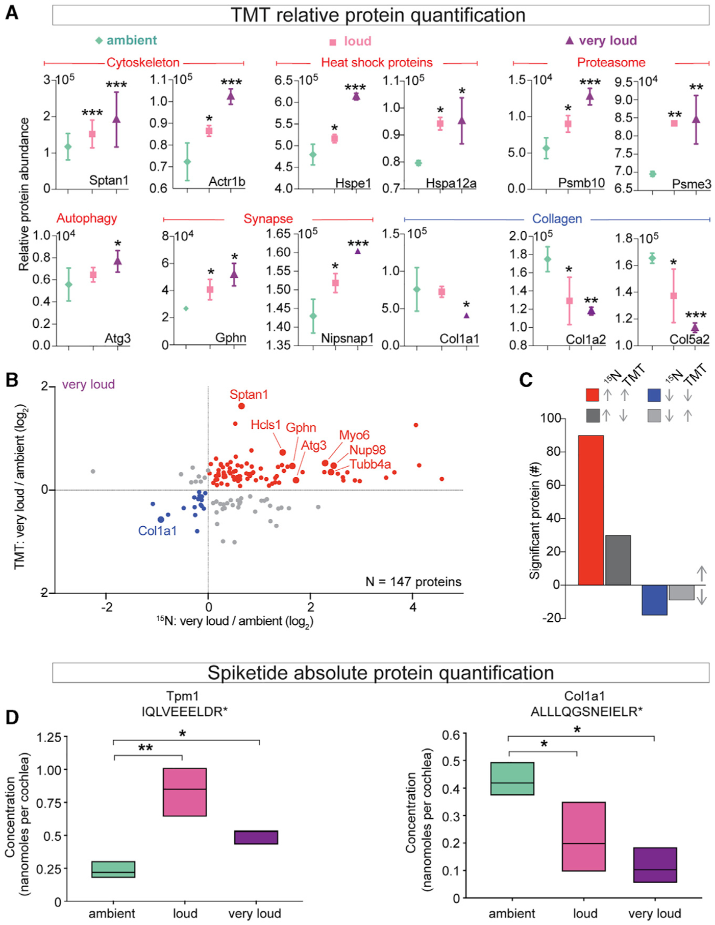
(A) Selected abundance plots for cytoskeletal, heat shock, proteasome, autophagy, synaptic, and collagen proteins with significantly elevated or reduced levels across noise exposures.
(B) Comparison of individual proteins with significantly altered FCs in very loud 15N and TMT datasets.
(C) Summary of protein FC trends for proteins significantly altered in both 15N and TMT very loud datasets. Of the 140 proteins found to be significantly altered by 15N, 66.6% of proteins with reduced levels and 75.0% of proteins with elevated levels were verified in TMT.
(D) Absolute quantification of Tpm1 (IQLVEEELDR) and Col1a1 (ALLLQGSNEIELR) based on the ratio of light and heavy (Arg+10) peptide-reconstructed MS1 chromatograms. The absolute abundance of Tpm1 was significantly elevated between loud versus ambient noise and very loud versus ambient noise (ambient = 0.233 ± 0.011, loud = 0.834 ± 0.0417, and very loud = 0.499 ± 0.0249, mean ± SD). The absolute abundance of Col1a1 was significantly reduced between loud versus ambient noise and very loud versus ambient noise (ambient = 0.428 ± 0.0214, loud = 0.215 ± 0.0107, and very loud = 0.114 ± 0.00570, mean ± SD, nanomoles per cochlea). n = 3 mice per noise exposure condition.
*p < 0.05, **p < 0.01, ***p < 0.001 by one-way ANOVA with Bonferroni correction (A); *p < 0.05, **p < 0.01 by one-way ANOVA with Holm-Sidak (D).