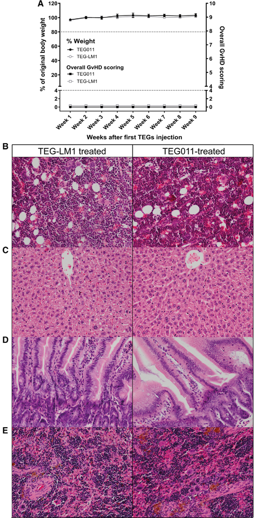
FIGURE 2. Weight loss, overall graft-versus-host disease (GvHD) scoring, and histopathology analysis of bone marrow and mouse vital organs (spleen, liver, intestine) of nontumor-bearing mice.
(A) Percentages of weight change measured weekly during study period for nontumor-bearing mice treated with TEG011 (filled black circle) and TEG-LM1 mock (open gray circle) tabulated on left Y-axis. A total of 20% weight loss from initial weight measured on day 1 were considered humane endpoint (HEP) and indicated by black tick line. Overall GvHD scoring was tabulated on right Y-axis for nontumor-bearing mice treated with TEG011 (filled black rectangle) and TEG-LM1 mock (open gray rectangle). Scoring was calculated based on following parameters: hunching, activity, fur texture, skin integrity, and diarrhea. Score range from 0 to 10 (see Supporting Information Table S1 for detail scoring system), where total overall score of 4 was considered HEP and indicated by black tick line. Score 0 depicts normal appearance for all GvHD parameters. Data represent mean ± SEM of all mice per group (n = 5 mice/group). (B) Representative photomicrographs H&E stained of mouse bone marrow from both TEG-LM1 mock (left panel) and TEG011-treated group (right panel). Magnification: 20×; (C) Representative photomicrographs for H&E stained of mouse liver for both TEG-LM1 mock (left) and TEG011-treated group (right) with apparent no histologic lesion. Magnification: 20×; (D) Representative pictures for H&E staining of mouse intestine for both TEG-LM1 mock (left) and TEG011-treated group (right) with apparent no histologic lesion. Magnification: 20×; (E) Representative photomicrographs for H&E stained of female mouse spleen for both TEG-LM1 mock (left) and TEG011-treated group (right) with a higher number of erythrocyte precursors and megakaryocytes. Magnification: 20×; Shown are representative photomicrographs from individual mice of both TEG011 and TEG-LM1 mock group (n = 5 mice/group) with no observable differences in overall histology features between treatment groups

