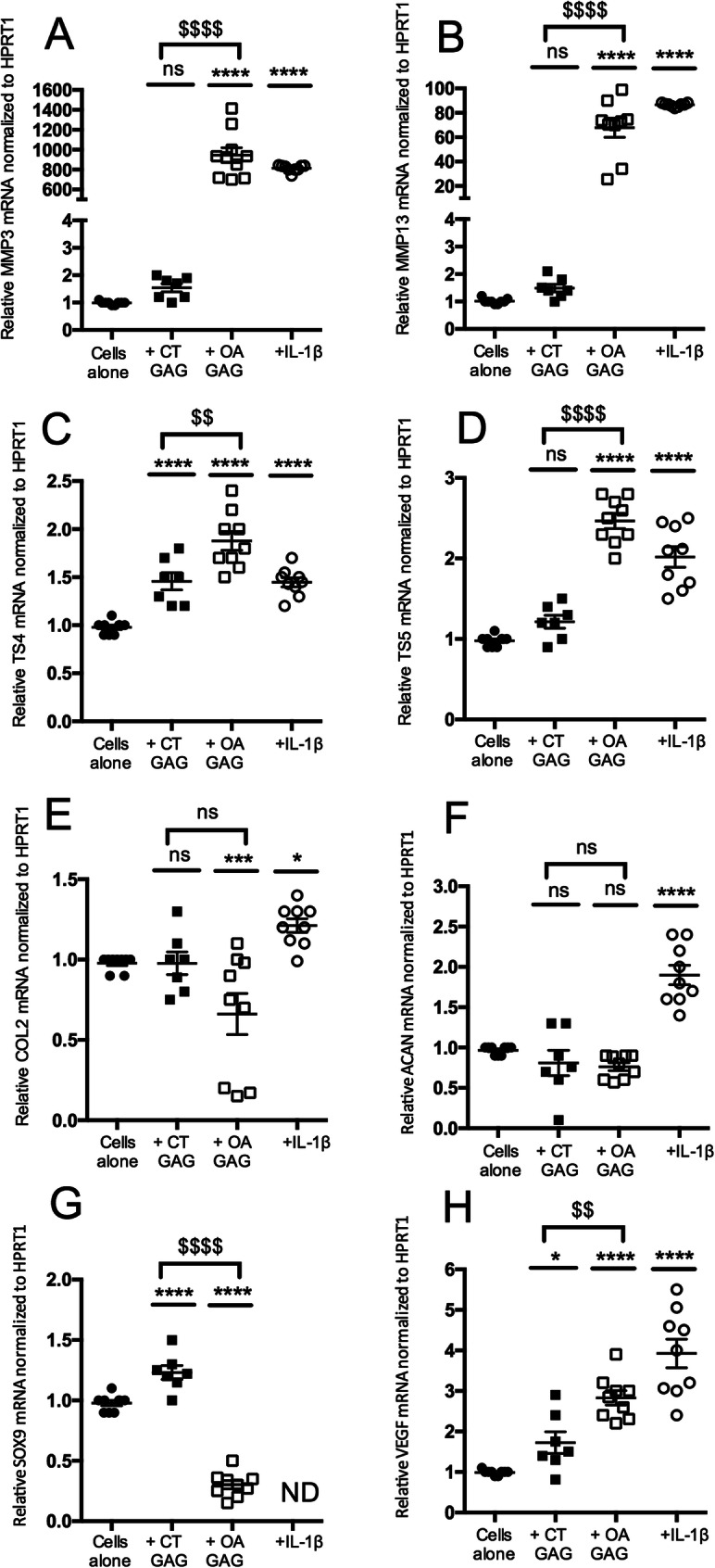
Fig. 4.

Effect of GAG from cartilages on murine chondrocyte phenotypes. Articular chondrocytes were grown in the absence (cells alone) or in the presence of total GAG (2.5 μg/mL) from the cartilage of control donors (CT GAG) or osteoarthritis patients (OA GAG), or in the presence of IL-1β (1 ng/mL). RQ-PCR analysis of the mRNA expression levels of catabolic markers, MMP3 (a), MMP13 (b), TS4 (c), and TS5 (d); anabolic markers, COL2 (e), ACAN (f) and SOX9 (g); and a hypertrophic marker, VEGF (h), reported to HPRT1 expression level (housekeeping gene) and compared to the basal condition (cells alone) defined as 1 (100%). Data is presented as the mean of 9 values obtained from 3 independent experiments in CT (n = 3 samples) and OA (n = 3 samples) cartilages. The statistical significance of the differences was determined using an ordinary one-way ANOVA test followed by pairwise comparisons using the Dunnett test compared to cells alone (*) and by a t test between CT GAG and OA GAG ($). Significant P values: *< 0.05, **< 0.01, ***< 0.001, ****< 0.0001, $$< 0.01, and $$$$< 0.0001
