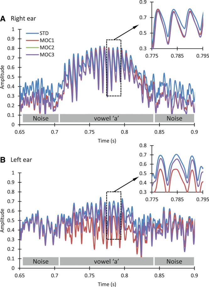
Fig. 2.

Zoomed-in view of the compressed envelopes for channel number 5 shown in Fig. 1. Each panel shows envelopes for the STD, MOC1, MOC2, and MOC3 strategies. Envelopes were identical for the MOC2 and MOC3, hence the overlap between corresponding traces. The gray rectangles near the abscissae depict periods when the noise or the vowel /a/ were present. A, Envelopes for the right ear. B, Envelopes for the left ear. The inset in each panel illustrates a zoomed-in view of the envelopes over the area depicted by the corresponding rectangle. MOC indicates medial olivocochlear; MOC1, original fast MOC strategy; MOC2, slower MOC strategy; MOC3, slower MOC strategy with comparatively greater contralateral inhibition in the lower-frequency than in the higher-frequency channels; STD, standard
