FIGURE 1.

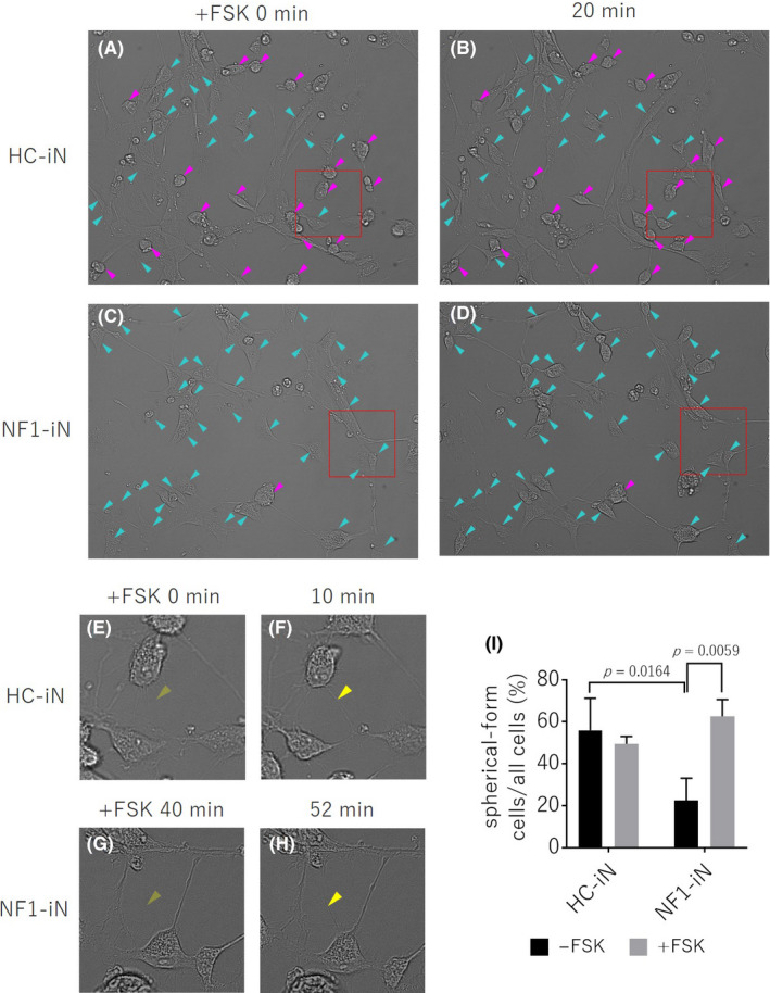
FIGURE (A‐D) Phase difference images of iN cells derived from healthy control (HC‐iN, A, B) and NF1 patient (NF1‐iN, C, D) after forskolin (FSK) treatment. Neuron‐like spherical‐form cells mainly appeared in HC‐iN cells (magenta arrow head). Flat cells with a thin cell contour obtain a dense cell contour only 20 minutes after forskolin treatment (cyan arrow head). (E‐H) Enlarged images of the part surrounded by the red frame on FIGURE A‐D. Forskolin appeared to enhanced neurite outgrowth in the iN cells (yellow arrow head). (I) The ratio of the number of neuronal‐like spherical‐form cells to the total number of cells. NF1‐iN cells in the absence of forskolin had a significantly lower percentage of the spherical‐form cells compared to HC‐iN cells (p = 0.0164, two‐way ANOVA / Tukey’s test, n = 3 each group). In the presence of forskolin, the spherical‐form cell morphology of NF1‐iN cells was significantly higher (p = 0.0059, two‐way ANOVA / Tukey’s test, n = 3 each group)
