
Figure 6.
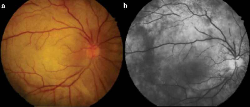
Case 4: Right eye (a) Colour fundus photograph showing segmental narrowing of the retinal arteries and a pale retina with central retinal artery occlusion. (b) Fluorescein angiogram demonstrating delayed filling of the central retinal artery with areas of patchy choroidal non-perfusion