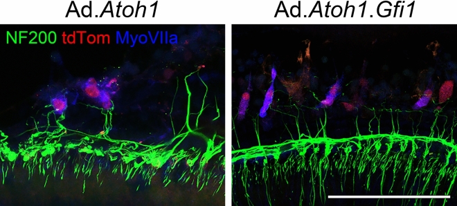
Figure 6.
Nerve fibers near regenerated HCLCs. Samples collected at 4 weeks after adenovirus (Ad) and DT-induced HC (HC) ablation. Spiral ganglion neuron (SGN) fibers were visualized by Neurofilament 200 (NF200) staining. Myosin VIIa (MyoVIIa) and tdTomato (tdTom) double-positive cells were associated with nerve fibers along the cell body. Both groups—adenovirus with Atoh1 (Ad.Atoh1) and with Atoh1 + Gfi1 (Ad.Gfi.Atoh1) showed similar association with SGNs. Representative images are shown from 4 biological replicates for each group. Scale bar = 100 μm.

