Fig. 2.
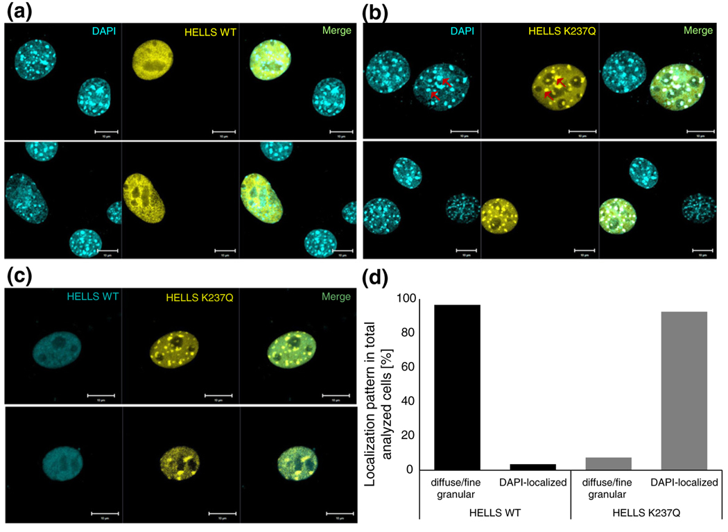
The wild-type and ATPase-deficient HELLS show distinct localization patterns in paraformaldehyde-fixed cells. (a) Representative confocal laser scanning microscopy images of DAPI-stained NIH 3T3 cells post-transient transfection with EYFP-fused wild-type HELLS. (b) Representative confocal laser scanning microscopy images of DAPI-stained NIH 3T3 cells post-transient transfection with EYFP-fused mutant HELLS. The red arrows indicate illustrative areas of co-localization between the protein and the DAPI-dense foci. (c) Representative confocal laser scanning microscopy images of NIH 3T3 cells post-transient co-transfection with ECFP-fused wild-type HELLS and EYFP-fused mutant HELLS. Scale bars correspond to 10 μm. (d) Quantification of localization patterns observed in wild-type or mutant HELLS transfected cells as illustratively shown in (a) and (b). We analyzed 29 and 27 cells for the wild-type and mutant HELLS transfected cells, respectively.

