
Figure 1.
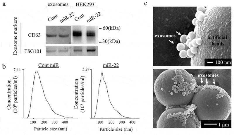
The exosome confirmation analysis. The exosomes were isolated from the culture medium of HEK293 cells transfected with either precursor miR-22 (miR-22) or control (cont miR). (a) Western blot analyses were performed to detect the exosomal marker proteins (CD63 and TSG101) in vesicles released by HEK293 cells. Representative examples of bands from three independent experiments are shown. (b) The particle size distributions and concentrations of exosomes were measured using a nanoparticle tracking system. (c) A representative image of exosomes using transmission electron microscopy