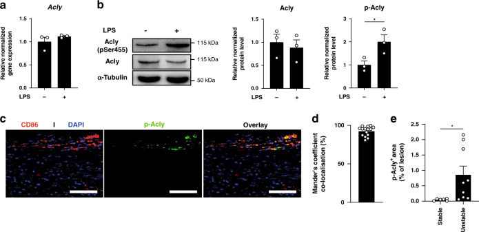
Fig. 1. Acly is phosphorylated in inflammatory conditions in vitro and in vivo.
a Relative normalized expression of Acly in unstimulated and LPS-stimulated macrophages. b Normalized protein levels from cell lysates of unstimulated and LPS-stimulated macrophages. Samples were immunoblotted with antibodies against Acly, phosphorylated Acly (p-Acly) and α-tubulin. Acly/p-Acly quantification on the blots derive from samples of the same experiment and gels/blots were processed in parallel. *P = 0.0441. c Representative immunohistochemical staining for macrophages (CD68) and p-ACLY in human plaques from 16 stable/unstable plaques. Scale bar represents 100 µm. d Quantification of colocalization, percentage of p-Acly+ macrophages overlapping with CD68+ area. e Quantification of p-Acly+ area in the lesion. *P = 0.0402. Values represent mean ± SEM (n = 3 technical replicates of three pooled mice a, n = 3 one representative image of three technical replicates of three pooled mice (b, western blot), n = 7/9 stable/unstable plaques d, e. *P < 0.05; by two-tailed Student’s t test (b, e). Source data are provided as a Source Data file (a–e).

