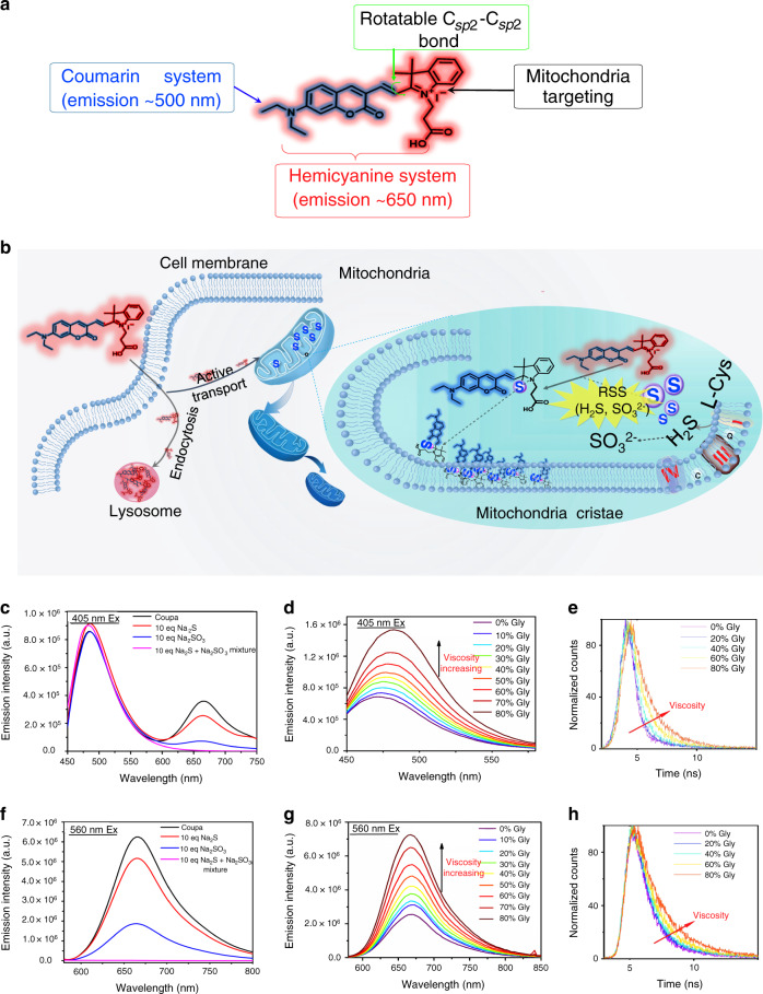
Fig. 1. Design and fluorescence characterization of Coupa.
a Chemical structure of Coupa and the role of each functional moieties. The structure possesses two ICT emission peaks—coumarin, which peaks at approximately 500 nm, and a hemicyanine system, which peaks at approximately 650 nm—that can target mitochondria via its positive charge and respond to viscosity via altering its – rotation. b Proposed mitochondria- and lysosome-staining mechanisms of Coupa. Coupa stains mitochondria by direct absorption, whereas lysosomal staining could result from active transport and endocytosis. Fluorescence spectra of Coupa were determined upon excitation at 405 nm (c and d) and 560 nm (f and g). c, f Fluorescence spectra determined with Na2S and Na2SO3 treatments, and Na2S and Na2SO3 completely quenched the red fluorescence (660 nm emission), leading to different Coupa emission with and without RSS. d, g Fluorescence spectra determined in media (glycerol/ methanol mixed solvent) with different viscosity; and e, h fluorescence decay profiles of Coupa in glycerol/methanol mixtures determined with picosecond pulsed excitation at 340 nm (e, Em at 535 nm) and 535 nm (h, Em at 670 nm).

