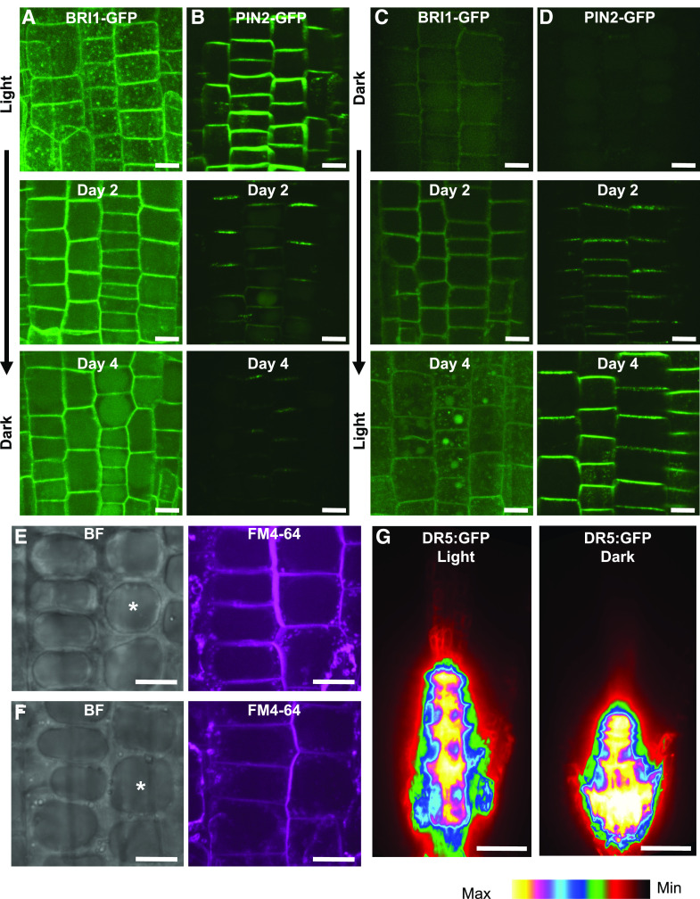
Figure 4.
PIN2 and BRI1 are not maintained at the PM in dark-grown plants. Representative images are confocal z-projections. A to D, BRI1-GFP (A and C) and PIN2-GFP (B and D) in light-grown (A and B) and dark-grown (C and D) division-zone cells of roots transferred to dark (A and B) and light (C and D) conditions and imaged at days 0, 2, and 4. Scale bars = 5 μm. E and F, Large central vacuoles (asterisks in brightfield [BF] images) in dark-grown BRI1-GFP (E) and PIN2-GFP (F) plants that are absent in light-grown samples. FM4-64 staining was used to show the PM and the tonoplast of the vacuole. Scale bars = 5 μm. G, DR5:GFP in light- and dark-grown root tips. Scale bars = 30 μm.

