Figure 4.
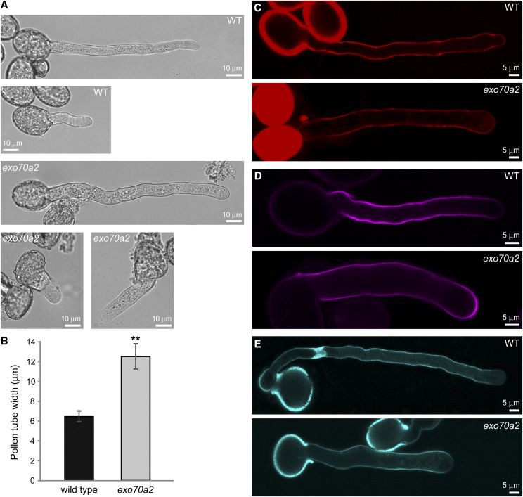
Morphology and cell wall deposition of exo70a2 and wild-type pollen tubes grown in vitro. A, Wild-type (WT) and exo70a2 pollen tubes 1.5 and 8 h after imbibition. B, Measurement of wild-type and exo70a2 pollen tube width (error bars represent the mean ± sd; n = 40). Asterisks indicate statistical difference using Student’s t test (P < 0.001). Pollen was collected from five different plants for each genotype, with two biological replicates. C to E, Visualization of pectins by propidium iodide staining (C), cellulose by Calcofluor White staining (D), and callose by aniline blue staining (E).

