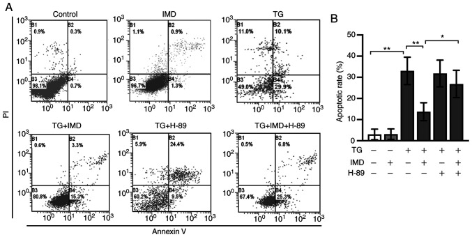
Figure 6.
IMD reduces endoplasmic reticulum stress-mediated apoptosis in cardiomyocytes. Neonatal rat cardiomyocytes were treated with TG in the presence or absence of IMD or H-89 for 24 h. Apoptotic rates were assessed using flow cytometry analysis. (A) Representative flow cytometric dot plots for each treatment group. Horizontal and vertical axis represent Annexin V and PI staining. Lower left quadrant, living cells; upper left quadrant, necrotic cells; and right quadrants, apoptotic cells. (B) Quantitative analysis of apoptotic cells. Data were analyzed with a student's t-test for two group comparison. Data are presented as the mean ± SD. n=3. *P<0.05 and **P<0.01. IMD, intermedin; PI, propidium iodide; TG, thapsigargin.

