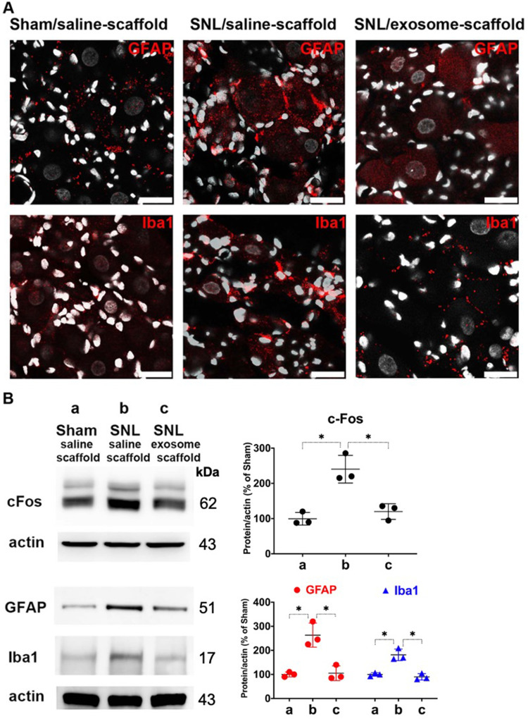
Figure 5.
Implantation of stem cell exosome-scaffold on ligated nerves attenuates L5/6 spinal nerve ligation (SNL)-induced neuron and glial activation in ipsilateral L5/6 dorsal root ganglion (DRG). (A) Representative immunofluorescent image showing SNL-induced GFAP (satellite glial cell marker) and Iba1 (macrophage marker) over-expression was attenuated by exosome-scaffold treatment on day 7 after surgery. The white spots are DAPI nuclear staining. Scale bar: 25 μm. (B) Left: representative Western blots of c-Fos (postsurgery day 1), GFAP and Iba1 (postsurgery day 7) of ipsilateral L5/6 DRG, actin used as internal control. Right: summarized graph depicting protein levels presented as raw data points and Mean ± SD, taking Sham/saline-scaffold group as 100% (*P<0.05, Kruskal–Wallis test followed by Dunn’s multiple pairwise comparisons test, n=3 in each group).

