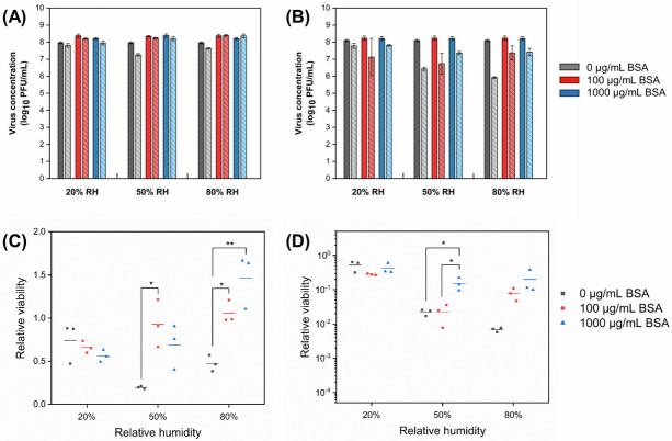
Fig 4.
Concentration of bacteriophages (A) MS2 and (B) Φ6 in droplets with different initial protein concentration before (dark bars) and after (light bars) 1 h exposure to low, intermediate, and high RH (mean ± s.d. of triplicates). Relative viability of (C) MS2 and (D) Φ6 after 1 h exposure (lines show the mean of triplicates). The number of virions in droplets at the start of the exposure experiments was 105−106 PFU.

