Figure 1.

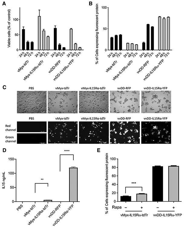
In vitro characterization of vMyx-tdTr, vMyx-IL15Rα-tdTr, vvDD-RFP, and vvDD-IL15Rα-YFP. A total of 2 × 10⁵ GL261WT cells were cultured per well of a 24-well plate, then infected with PBS, vMyx-tdTr, vMyx-IL15Rα-tdTr, vvDD-RFP, or vvDD-IL15Rα-YFP at MOI 5 for 24, 48, or 72 hours. A, Viable cell counts. B, Flow cytometry analysis of tdTomato Red (vMyx-tdTr and vMyx-IL15Rα-tdTr), RFP (vvDD-RFP), and YFP (vvDD-IL15Rα-YFP). Mean values and SEM are shown. C, Brightfield and fluorescence images of GL261 WT cells were taken 48 hours after virus infection. Red channel: tdTomato Red (vMyx-tdTr and vMyx-IL15Rα-tdTr), RFP (vvDD-RFP); green channel: YFP (vvDD-IL15Rα-YFP). Scale bar, 100 μm. D, Supernatants of cells from each treatment were collected 48 hours after virus infection. The production of IL15Rα-IL15 was measured by ELISA. Mean ELISA values and SEM are shown. There was a significant increase in IL15Rα-IL15 expression in the supernatants of vMyx-IL15Rα-tdTr or vvDD-IL15Rα-YFP–treated cells compared with vMyx-tdTr or vvDD-RFP–treated cells. P values: **, < 0.01; ****, < 0.0001. E, GL261 WT cells were treated with or without 100 nmol/L rapamycin 2 hours prior to the 1-hour inoculation of vMyx-IL15Rα-tdTr or vvDD-IL15Rα-YFP at MOI 5. Cells were fixed and analyzed for tdTomato red (vMyx-IL15Rα-tdTr) or YFP (vvDD-IL15Rα-YFP) expression by flow cytometry at 48 hours p.i. Mean ± SEM, n = 3, P value: ***, < 0.001.
