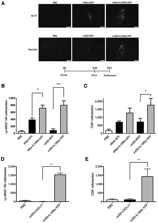
Figure 2.

In vivo characterization of vMyx-tdTr, vMyx-IL15Rα-tdTr, vvDD-RFP, and vvDD-IL15Rα-YFP. C57BL/6J mice were implanted with GL261 NS cells intracranially followed 10 days later by injectionwith PBS, vMyx-tdTr, vMyx-IL15Rα-tdTr, vvDD-RFP, or vvDD-IL15Rα-YFPfor A-C, and PBS, vvDD-CXCL11, or vvDD-IL15Rα-YFP for D and E (n = 3;2 × 10⁶ pfu intratumoral). Mice were euthanized 5 days after the virus treatment and tumor sections were analyzed for presence of the viruses, NKcells, and CD8+ T cells by immunostaining for M-T7 (a myxoma-encoded protein), vaccinia virus, Ly-49G2 (4D11 antibody) for NK cells and CD8, respectively. Representative tumor sections are shown. A, Staining for vMyx or vvDDvirus in tumors. Scale bar, 200 μm. B, Numberof Ly-49G2+ NKcells per tumor section for each condition, mean values and SEM are shown. One-way ANOVA showed significant increase in NK cell accumulation in vMyx-IL15Rα-tdTr or vvDD-IL15Rα-YFP–treated tumors compared with vMyx-tdTr or vvDD-RFP treatments. P values: **, < 0.01; ***, < 0.001. C, Number of CD8+ cells per tumor section for each condition. Mean values and SEM are shown. One-way ANOVA showed significant increase in CD8+ cell accumulation in vvDD-IL15Rα-YFP–treated tumors compared with vvDD-RFP treatment. P value: *, < 0.05. D, Number of Ly-49G2+ NK cells per tumor section for each condition. Mean values and SEM are shown. One-way ANOVA showed significant increase in NK-cell accumulation in vvDD-IL15Rα-YFP–treated tumors compared with both vvDD-CXCL11 and PBS treatments. P value: **, < 0.01. E, Number of CD8+ cells per tumor section for each condition. Mean values and SEM are shown. One-way ANOVA showed significant increase in CD8+ cell accumulation in vvDD-IL15Rα-YFP–treated tumors compared with both vvDD-CXCL11 and PBS treatments. P value: **, < 0.01.
