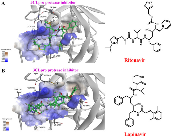
Figure 11.
Molecular docking of ritonavir and lopinavir binding to the 3CLpro. (A) The right panel shows the structure of ritonavir, the left panel shows molecular docking simulation using Discovery Studio 2020. (B) The right panel is the structure of lopinavir, the left panel shows molecular docking simulation. The structures of the drugs are presented using a stick model. Carbon atoms are coloured green. 3CLpro, 3-chymotrypsin-like cysteine protease.

