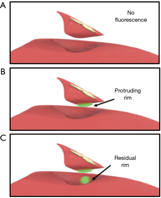
Figure 2.

A computer-aided design of the liver showcasing three different scenarios. (A) No fluorescent signal produced by either the liver’s wound bed or the resection specimen. (B) Fluorescent rim protruding trough the liver tissue of the resection specimen indicating a potential tumor-positive resection margin and (C) bright fluorescent signal in both the wound bed and protruding trough the liver tissue of the resection specimen (potential resection with tumor-positive margin).
