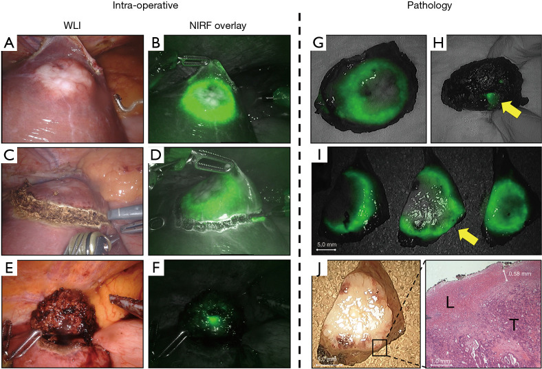
Figure 4.
Correlation between in vivo and ex vivo NIRF imaging. (A) Resection of a CRLM in liver segment IVb in normal white light and, (B) with NIRF imaging. (C) Too close proximity of the demarcation line to the (D) fluorescent rim might result in a resection with tumor-positive margin. (E) Suspected resection with tumor-positive margin using WLI and (F) confirmed with NIRF. Ex vivo NIRF imaging of the (G) front of the lesion and (H) from the back show the same protruding rim. (I) Gross sectioning reveals a potential resection with tumor-positive margin, indicated by the yellow arrow. Which is confirmed by NIRF imaging. Later histopathological assessment of the suspected tumor-margin in (J) is confirmed by H&E staining. WLI, white light imaging; NIRF, near-infrared fluorescence; H&E, haematoxylin and eosin; L, normal liver tissue; T, tumor tissue.

