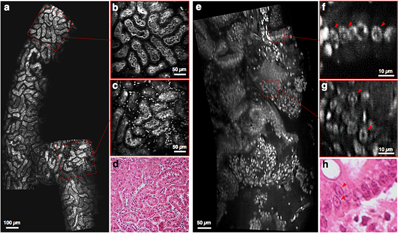
Fig. 5.

Video mosaicking of fluorescently label fresh mouse tissues imaged at 16 fps (depth of imaging ~50 μm). (a-c) Sub-tubular structures in the cortex of a fresh mouse kidney are clearly visualized, and (d) agree with corresponding H&E histology. (e-g) Another example in a mouse colon shows that sub-nuclear structures are distinguishable, with good agreement with (h) corresponding H&E histology. [See supplemental visualizations for the corresponding video clips]
