
Fig. 1.
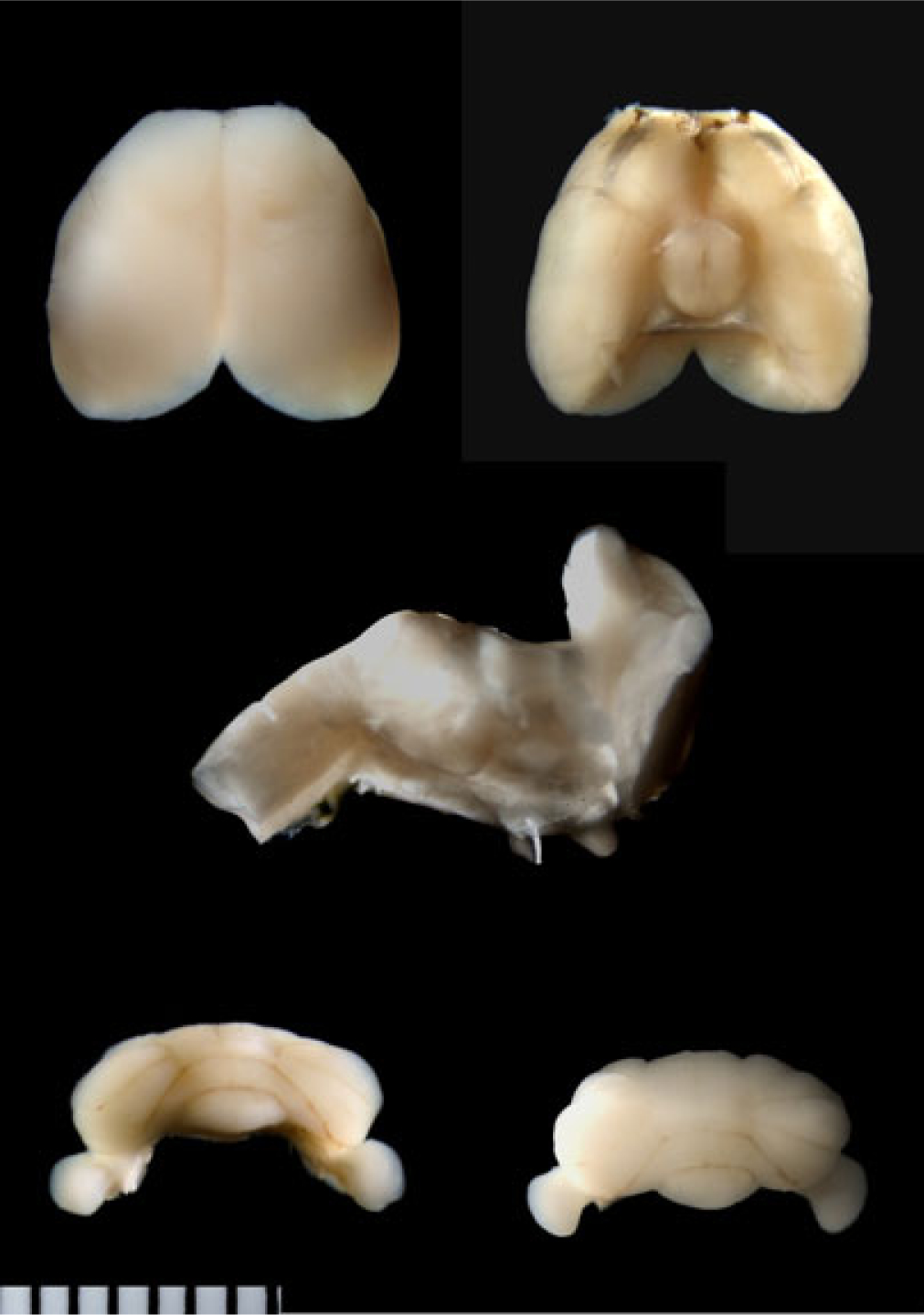
The mouse brain. The 2 top photos show dorsal and ventral views of the mouse cerebral cortex prior to the midsagittal cut to expose the corpus callosum. The olfactory appendages are cut flush at the level of the anterior end of the cerebral cortex. A cut mark (that separated the brainstem) is visible in the ventral view. The middle photo shows a sagittal view of the cut-away brainstem. Here, the rostral cut plane is visible, indicating the cerebral cortex was dissected free just rostral to the superior colliculi on the dorsal surface of the brainstem and just caudal to the pons on the ventral surface of the brainstem. The bottom photos show anterior and posterior views of the cerebellum. Each division of the marker is 0.6 mm.